Structural dynamics in proteins induced by and probed with X-ray free-electron laser pulses.
Nass, K., Gorel, A., Abdullah, M.M., V Martin, A., Kloos, M., Marinelli, A., Aquila, A., Barends, T.R.M., Decker, F.J., Bruce Doak, R., Foucar, L., Hartmann, E., Hilpert, M., Hunter, M.S., Jurek, Z., Koglin, J.E., Kozlov, A., Lutman, A.A., Kovacs, G.N., Roome, C.M., Shoeman, R.L., Santra, R., Quiney, H.M., Ziaja, B., Boutet, S., Schlichting, I.(2020) Nat Commun 11: 1814-1814
- PubMed: 32286284 Search on PubMedSearch on PubMed Central
- DOI: https://doi.org/10.1038/s41467-020-15610-4
- Primary Citation Related Structures:
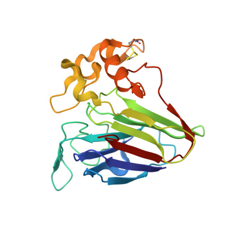
6SR0, 6SR1, 6SR2, 6SR3, 6SR4, 6SR5, 6SRJ, 6SRK, 6SRL, 6SRO, 6SRP, 6SRQ - PubMed Abstract:
X-ray free-electron lasers (XFELs) enable crystallographic structure determination beyond the limitations imposed upon synchrotron measurements by radiation damage. The need for very short XFEL pulses is relieved through gating of Bragg diffraction by loss of crystalline order as damage progresses, but not if ionization events are spatially non-uniform due to underlying elemental distributions, as in biological samples. Indeed, correlated movements of iron and sulfur ions were observed in XFEL-irradiated ferredoxin microcrystals using unusually long pulses of 80 fs. Here, we report a femtosecond time-resolved X-ray pump/X-ray probe experiment on protein nanocrystals. We observe changes in the protein backbone and aromatic residues as well as disulfide bridges. Simulations show that the latter's correlated structural dynamics are much slower than expected for the predicted high atomic charge states due to significant impact of ion caging and plasma electron screening. This indicates that dense-environment effects can strongly affect local radiation damage-induced structural dynamics.
- Max-Planck-Institut für Medizinische Forschung, Jahnstraße 29, 69120, Heidelberg, Germany.
Organizational Affiliation: