6SNN | pdb_00006snn

Crystal structure of cAMP-dependent protein kinase A (CHO PKA) in complex with benzoic acid


Experimental Data Snapshot

  • Method: X-RAY DIFFRACTION
  • Resolution: 1.82 Å
  • R-Value Free: 
    0.209 (Depositor), 0.211 (DCC) 
  • R-Value Work: 
    0.173 (Depositor), 0.176 (DCC) 

wwPDB Validation 3D Report Full Report

Validation slider image for 6SNN

Ligand Structure Quality Assessment 


This is version 1.3 of the entry. See complete history

Literature

Fragment Binding to Kinase Hinge: If Charge Distribution and Local pK a Shifts Mislead Popular Bioisosterism Concepts.

Oebbeke, M.Siefker, C.Wagner, B.Heine, A.Klebe, G.

(2021) Angew Chem Int Ed Engl 60: 252-258

  • DOI: https://doi.org/10.1002/anie.202011295
  • Primary Citation Related Structures: 
    5N33, 5N3C, 5N3D, 5N3E, 5N3H, 5N3J, 5N3Q, 5N3S, 6SNN, 6SNX, 6SOX, 6SPM, 6SPS, 6SPU, 6SPY, 6YPS, 6Z08, 6Z44, 6ZN0

  • PubMed Abstract: 

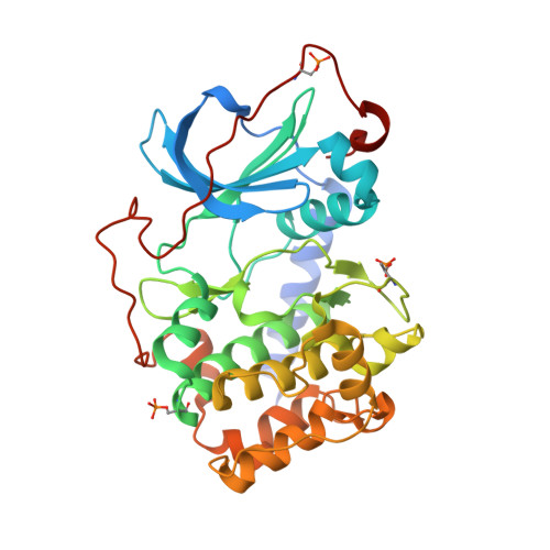
    Medicinal-chemistry optimization follows strategies replacing functional groups and attaching larger substituents at a promising lead scaffold. Well-established bioisosterism rules are considered, however, it is difficult to estimate whether the introduced modifications really match the required properties at a binding site. The electron density distribution and pK a values are modulated influencing protonation states and bioavailability. Considering the adjacent H-bond donor/acceptor pattern of the hinge binding motif in a kinase, we studied by crystallography a set of fragments to map the required interaction pattern. Unexpectedly, benzoic acid and benzamidine, decorated with the correct substituents, are totally bioisosteric just as carboxamide and phenolic OH. A mono-dentate pyridine nitrogen out-performs bi-dentate functionalities. The importance of correctly designing pK a values of attached functional groups by additional substituents at the parent scaffold is rendered prominent.


  • Organizational Affiliation
    • Philipps Universität Marburg, Institut für Pharmazeutische Chemie, Marbacher Weg 6, 35032, Marburg, Germany.

Macromolecule Content 

  • Total Structure Weight: 41.16 kDa 
  • Atom Count: 3,079 
  • Modeled Residue Count: 339 
  • Deposited Residue Count: 351 
  • Unique protein chains: 1

Macromolecules

Find similar proteins by:|  3D Structure
Entity ID: 1
MoleculeChains  Sequence LengthOrganismDetailsImage
cAMP-dependent protein kinase catalytic subunit alpha351Cricetulus griseusMutation(s): 0 
Gene Names: PRKACA
EC: 2.7.11.11
UniProt
Find proteins for P25321 (Cricetulus griseus)
Explore P25321 
Go to UniProtKB:  P25321
Entity Groups
Sequence Clusters30% Identity50% Identity70% Identity90% Identity95% Identity100% Identity
UniProt GroupP25321
Sequence Annotations
Expand
Reference Sequence

Small Molecules

Modified Residues  2 Unique
IDChains TypeFormula2D DiagramParent
SEP
Query on SEP
A
L-PEPTIDE LINKINGC3 H8 N O6 PSER
TPO
Query on TPO
A
L-PEPTIDE LINKINGC4 H10 N O6 PTHR

Experimental Data & Validation

Experimental Data

  • Method: X-RAY DIFFRACTION
  • Resolution: 1.82 Å
  • R-Value Free:  0.209 (Depositor), 0.211 (DCC) 
  • R-Value Work:  0.173 (Depositor), 0.176 (DCC) 
Space Group: P 21 21 21
Unit Cell:
Length ( Å )Angle ( ˚ )
a = 52.278α = 90
b = 71.526β = 90
c = 98.435γ = 90
Software Package:
Software NamePurpose
PHASERphasing
Cootmodel building
PHENIXrefinement
XDSdata reduction
XDSdata scaling

Structure Validation

View Full Validation Report


Ligand Structure Quality Assessment 


Entry History 

Deposition Data

Revision History  (Full details and data files)

  • Version 1.0: 2020-09-09
    Type: Initial release
  • Version 1.1: 2020-12-09
    Changes: Database references
  • Version 1.2: 2020-12-30
    Changes: Database references
  • Version 1.3: 2024-10-23
    Changes: Data collection, Database references, Structure summary