6SI7 | pdb_00006si7

Structure of the curli secretion-assembly complex CsgG:CsgF


Experimental Data Snapshot

  • Method: ELECTRON MICROSCOPY
  • Resolution: 3.40 Å
  • Aggregation State: PARTICLE 
  • Reconstruction Method: SINGLE PARTICLE 

Starting Model: experimental
View more details

wwPDB Validation 3D Report Full Report

Validation slider image for 6SI7

This is version 1.4 of the entry. See complete history

Literature

A dual-constriction biological nanopore resolves homonucleotide sequences with high fidelity.

Van der Verren, S.E.Van Gerven, N.Jonckheere, W.Hambley, R.Singh, P.Kilgour, J.Jordan, M.Wallace, E.J.Jayasinghe, L.Remaut, H.

(2020) Nat Biotechnol 38: 1415-1420

  • DOI: https://doi.org/10.1038/s41587-020-0570-8
  • Primary Citation Related Structures: 
    6SI7

  • PubMed Abstract: 

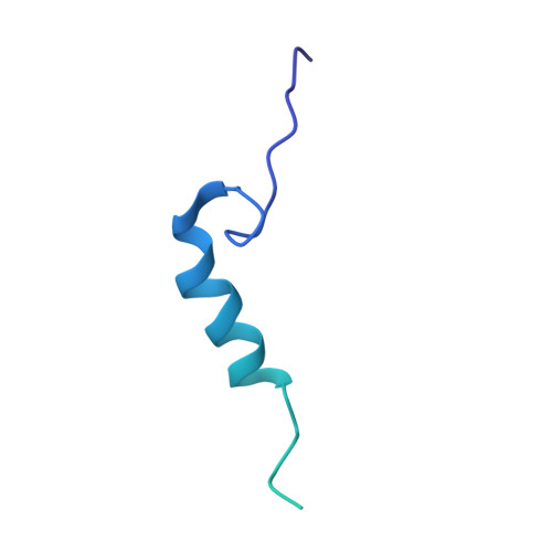
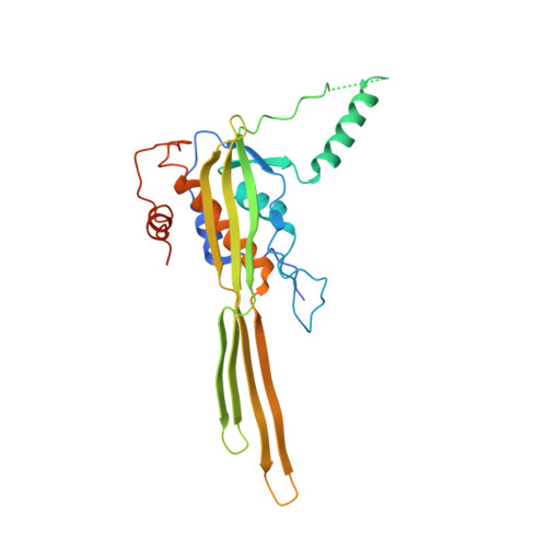
    Single-molecule long-read DNA sequencing with biological nanopores is fast and high-throughput but suffers reduced accuracy in homonucleotide stretches. We now combine the CsgG nanopore with the 35-residue N-terminal region of its extracellular interaction partner CsgF to produce a dual-constriction pore with improved signal and base-calling accuracy for homopolymer regions. The electron cryo-microscopy structure of CsgG in complex with full-length CsgF shows that the 33 N-terminal residues of CsgF bind inside the β-barrel of the pore, forming a defined second constriction. In complexes of CsgG bound to a 35-residue CsgF constriction peptide, the second constriction is separated from the primary constriction by ~25 Å. We find that both constrictions contribute to electrical signal modulation during single-stranded DNA translocation. DNA sequencing using a prototype CsgG-CsgF protein pore with two constrictions improved single-read accuracy by 25 to 70% in homopolymers up to 9 nucleotides long.


  • Organizational Affiliation
    • Structural Biology Brussels, Vrije Universiteit Brussel, Brussels, Belgium.

Macromolecule Content 

  • Total Structure Weight: 394.7 kDa 
  • Atom Count: 19,476 
  • Modeled Residue Count: 2,502 
  • Deposited Residue Count: 3,573 
  • Unique protein chains: 2

Macromolecules

Find similar proteins by:|  3D Structure
Entity ID: 1
MoleculeChains  Sequence LengthOrganismDetailsImage
Curli production assembly/transport component CsgF125Escherichia coliMutation(s): 0 
Gene Names: csgFb1038JW1021
Membrane Entity: Yes 
UniProt
Find proteins for P0AE98 (Escherichia coli (strain K12))
Explore P0AE98 
Go to UniProtKB:  P0AE98
Entity Groups
Sequence Clusters30% Identity50% Identity70% Identity90% Identity95% Identity100% Identity
UniProt GroupP0AE98
Sequence Annotations
Expand
Reference Sequence
Find similar proteins by:|  3D Structure
Entity ID: 2
MoleculeChains  Sequence LengthOrganismDetailsImage
Curli production assembly/transport component CsgG272Escherichia coliMutation(s): 0 
Gene Names: csgGb1037JW1020
Membrane Entity: Yes 
UniProt
Find proteins for P0AEA2 (Escherichia coli (strain K12))
Explore P0AEA2 
Go to UniProtKB:  P0AEA2
Entity Groups
Sequence Clusters30% Identity50% Identity70% Identity90% Identity95% Identity100% Identity
UniProt GroupP0AEA2
Sequence Annotations
Expand
Reference Sequence

Experimental Data & Validation

Experimental Data

  • Method: ELECTRON MICROSCOPY
  • Resolution: 3.40 Å
  • Aggregation State: PARTICLE 
  • Reconstruction Method: SINGLE PARTICLE 
EM Software:
TaskSoftware PackageVersion
RECONSTRUCTIONRELION2.0
MODEL REFINEMENTPHENIX1.14

Structure Validation

View Full Validation Report


Entry History 

& Funding Information

Deposition Data


Funding OrganizationLocationGrant Number
European Research CouncilBelgium649082
Research Foundation - FlandersBelgium--

Revision History  (Full details and data files)

  • Version 1.0: 2020-06-24
    Type: Initial release
  • Version 1.1: 2020-07-15
    Changes: Database references
  • Version 1.2: 2020-11-18
    Changes: Structure summary
  • Version 1.3: 2020-12-16
    Changes: Database references
  • Version 1.4: 2024-05-22
    Changes: Data collection, Database references, Refinement description