Protein-mediated disproportionation of Au(i): insights from the structures of adducts of Au(iii) compounds bearing N,N-pyridylbenzimidazole derivatives with lysozyme.
Ferraro, G., Giorgio, A., Mansour, A.M., Merlino, A.(2019) Dalton Trans 48: 14027-14035
- PubMed: 31490509 Search on PubMed
- DOI: https://doi.org/10.1039/c9dt02729g
- Primary Citation Related Structures:
6SET, 6SEU, 6SEW, 6SEX, 6SEZ - PubMed Abstract:
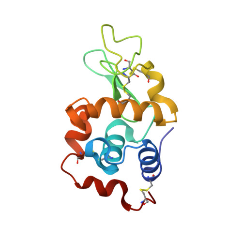
Au(iii) compounds bearing N,N-pyridylbenzimidazole derivatives with the ethyl (1) or propyl sulfonate (2) appendage react with the model protein hen egg white lysozyme (HEWL), forming adducts with different gold-containing fragments. The conformation of the enzyme, the exact gold binding sites and the oxidation state of Au in the adducts are unknown. Here we report a structural study on the reaction of 1 and 2 with HEWL in solution and solid state. In agreement with previously reported electrospray ionization mass spectra, the compounds degrade in their interaction with the protein. In the structure derived from HEWL crystals exposed to 1 for less than one day, three Au binding sites were identified: Au(i) ions are bound to the side chain of His15 and to the side chains of His15 and Asn93. The third gold centre is buried in the hydrophobic pocket of the protein via the binding to the side chain of Met105 and the trapping between the side chains of Trp28, Trp108 and Trp111. In a second crystal fished three hours later from the same drop, only one Au ion, probably in the +1 oxidation state, is observed; it binds the protein close to the side chains of Asn93 and His15. After three days of soaking, the colour of HEWL crystals obtained in the presence of 1 turned violet. In these crystals, anomalous signals attributable to Au are found on the protein surface; gold atoms are not directly coordinated to residue side chains. Longer exposure of HEWL crystals to 1 produces gold-free crystals. In the adduct of HEWL exposed to 2 for one day, three Au(i) ions are detected close to the side chains of both Asn93 and His15, the side chain of His15 and that of Met105. Longer exposure of HEWL crystals to 2 affords gold-free crystals. These structural data and those of the other protein/gold adducts available at the Protein Data Bank suggest that the reduction of Au(iii) into Au(i) is the basis of the mechanism of action of the biologically active gold(iii) compounds. Besides, Au(i) ions can undergo disproportionation into Au(iii) and Au(0) that can diffuse away from the protein crystals.
- Department of Chemistry "Ugo Schiff", University of Florence, via della Lastruccia, 3-13, 50019, Sesto Fiorentino, Florence, Italy.
Organizational Affiliation: