


Kinase Scaffold Repurposing in the Public Domain
Sorrell, F.J., Henderson, S.H., Elkins, J.M., Ward, S.To be published.
Experimental Data Snapshot
Starting Model: experimental
View more details
Entity ID: 1 | |||||
|---|---|---|---|---|---|
| Molecule | Chains | Sequence Length | Organism | Details | Image |
| Dual specificity tyrosine-phosphorylation-regulated kinase 1A | 361 | Homo sapiens | Mutation(s): 0 Gene Names: DYRK1A, DYRK, MNB, MNBH EC: 2.7.12.1 (PDB Primary Data), 2.7.11.23 (UniProt) | 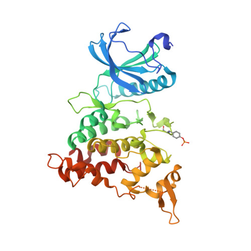 | |
UniProt & NIH Common Fund Data Resources | |||||
PHAROS: Q13627 GTEx: ENSG00000157540 | |||||
Entity Groups | |||||
| Sequence Clusters | 30% Identity50% Identity70% Identity90% Identity95% Identity100% Identity | ||||
| UniProt Group | Q13627 | ||||
Sequence AnnotationsExpand | |||||
Reference Sequence | |||||
| Ligands 4 Unique | |||||
|---|---|---|---|---|---|
| ID | Chains | Name / Formula / InChI Key | 2D Diagram | 3D Interactions | |
| KRK (Subject of Investigation/LOI) Download:Ideal Coordinates CCD File | B [auth A] | 3-[2-[(3~{S})-3-fluoranylpyrrolidin-1-yl]pyrimidin-4-yl]pyrazolo[1,5-b]pyridazine C14 H13 F N6 CTKSUQYBSSCMMB-JTQLQIEISA-N |  | ||
| SO4 Download:Ideal Coordinates CCD File | J [auth A], K [auth A], L [auth A], M [auth A] | SULFATE ION O4 S QAOWNCQODCNURD-UHFFFAOYSA-L |  | ||
| DMS Download:Ideal Coordinates CCD File | G [auth A] | DIMETHYL SULFOXIDE C2 H6 O S IAZDPXIOMUYVGZ-UHFFFAOYSA-N |  | ||
| EDO Download:Ideal Coordinates CCD File | C [auth A] D [auth A] E [auth A] F [auth A] H [auth A] | 1,2-ETHANEDIOL C2 H6 O2 LYCAIKOWRPUZTN-UHFFFAOYSA-N |  | ||
| Modified Residues 1 Unique | |||||
|---|---|---|---|---|---|
| ID | Chains | Type | Formula | 2D Diagram | Parent |
| PTR Query on PTR | A | L-PEPTIDE LINKING | C9 H12 N O6 P |  | TYR |
| Length ( Å ) | Angle ( ˚ ) |
|---|---|
| a = 99.202 | α = 90 |
| b = 69.671 | β = 117.54 |
| c = 67.44 | γ = 90 |
| Software Name | Purpose |
|---|---|
| Aimless | data scaling |
| PHASER | phasing |
| PHENIX | refinement |
| PDB_EXTRACT | data extraction |
| MOSFLM | data reduction |


