


Crystal structure of Escherichia coli Glyoxalase II
Skorupskaite, A., McDonough, M.A., Brem, J., Schofield, C.J.To be published.
Experimental Data Snapshot
Starting Model: experimental
View more details
wwPDB Validation 3D Report Full Report

Entity ID: 1 | |||||
|---|---|---|---|---|---|
| Molecule | Chains | Sequence Length | Organism | Details | Image |
| Hydroxyacylglutathione hydrolase GloB | 251 | Escherichia coli K-12 | Mutation(s): 0 Gene Names: gloB, yafR, b0212, JW0202 EC: 3.1.2.6 | 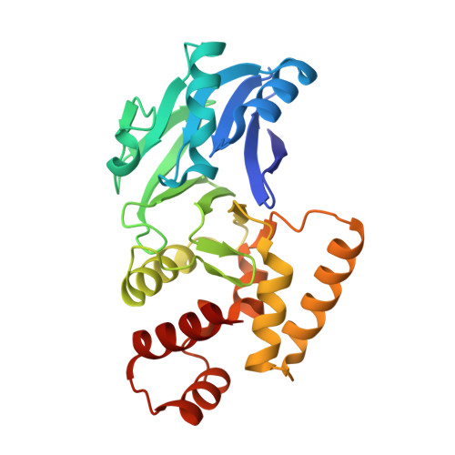 | |
UniProt | |||||
Entity Groups | |||||
| Sequence Clusters | 30% Identity50% Identity70% Identity90% Identity95% Identity100% Identity | ||||
| UniProt Group | P0AC84 | ||||
Sequence AnnotationsExpand | |||||
Reference Sequence | |||||
| Ligands 3 Unique | |||||
|---|---|---|---|---|---|
| ID | Chains | Name / Formula / InChI Key | 2D Diagram | 3D Interactions | |
| EPE Download:Ideal Coordinates CCD File | E [auth A] | 4-(2-HYDROXYETHYL)-1-PIPERAZINE ETHANESULFONIC ACID C8 H18 N2 O4 S JKMHFZQWWAIEOD-UHFFFAOYSA-N |  | ||
| TLA Download:Ideal Coordinates CCD File | D [auth A] | L(+)-TARTARIC ACID C4 H6 O6 FEWJPZIEWOKRBE-JCYAYHJZSA-N |  | ||
| ZN Download:Ideal Coordinates CCD File | B [auth A], C [auth A] | ZINC ION Zn PTFCDOFLOPIGGS-UHFFFAOYSA-N |  | ||
| Length ( Å ) | Angle ( ˚ ) |
|---|---|
| a = 39.053 | α = 90 |
| b = 43.655 | β = 90 |
| c = 159.067 | γ = 90 |
| Software Name | Purpose |
|---|---|
| PHENIX | refinement |
| Aimless | data scaling |
| PHASER | phasing |
| PDB_EXTRACT | data extraction |
| XDS | data reduction |
| Funding Organization | Location | Grant Number |
|---|---|---|
| Biotechnology and Biological Sciences Research Council | United Kingdom | BB/M011224/1 |


