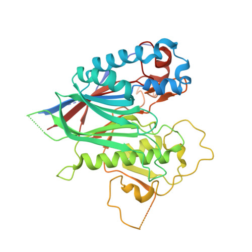
6RVZ

Crystal structure of ANGEL2, a 2',3'-cyclic phosphatase, in complex with adenosine-2',3'-vanadate


Experimental Data Snapshot

  • Method: X-RAY DIFFRACTION
  • Resolution: 2.10 Å
  • R-Value Free: 0.211 
  • R-Value Work: 0.188 
  • R-Value Observed: 0.189 

Starting Model: experimental
View more details

wwPDB Validation   3D Report Full Report


Ligand Structure Quality Assessment 


This is version 1.2 of the entry. See complete history


Literature

ANGEL2 is a member of the CCR4 family of deadenylases with 2',3'-cyclic phosphatase activity.

Pinto, P.H.Kroupova, A.Schleiffer, A.Mechtler, K.Jinek, M.Weitzer, S.Martinez, J.

(2020) Science 369: 524-530

  • DOI: https://doi.org/10.1126/science.aba9763
  • Primary Citation of Related Structures:  
    6RVZ, 6RW0

  • PubMed Abstract: 

    RNA molecules are frequently modified with a terminal 2',3'-cyclic phosphate group as a result of endonuclease cleavage, exonuclease trimming, or de novo synthesis. During pre-transfer RNA (tRNA) and unconventional messenger RNA (mRNA) splicing, 2',3'-cyclic phosphates are substrates of the tRNA ligase complex, and their removal is critical for recycling of tRNAs upon ribosome stalling. We identified the predicted deadenylase angel homolog 2 (ANGEL2) as a human phosphatase that converts 2',3'-cyclic phosphates into 2',3'-OH nucleotides. We analyzed ANGEL2's substrate preference, structure, and reaction mechanism. Perturbing ANGEL2 expression affected the efficiency of pre-tRNA processing, X-box-binding protein 1 ( XBP1 ) mRNA splicing during the unfolded protein response, and tRNA nucleotidyltransferase 1 (TRNT1)-mediated CCA addition onto tRNAs. Our results indicate that ANGEL2 is involved in RNA pathways that rely on the ligation or hydrolysis of 2',3'-cyclic phosphates.


  • Organizational Affiliation

    Max Perutz Labs, Medical University of Vienna, Vienna BioCenter, 1030 Vienna, Austria.


Macromolecules
Find similar proteins by:  (by identity cutoff)  |  3D Structure
Entity ID: 1
MoleculeChains Sequence LengthOrganismDetailsImage
Protein angel homolog 2429Homo sapiensMutation(s): 0 
Gene Names: ANGEL2KIAA0759L
UniProt & NIH Common Fund Data Resources
Find proteins for Q5VTE6 (Homo sapiens)
Explore Q5VTE6 
Go to UniProtKB:  Q5VTE6
PHAROS:  Q5VTE6
GTEx:  ENSG00000174606 
Entity Groups  
Sequence Clusters30% Identity50% Identity70% Identity90% Identity95% Identity100% Identity
UniProt GroupQ5VTE6
Sequence Annotations
Expand
  • Reference Sequence
Small Molecules
Ligands 4 Unique
IDChains Name / Formula / InChI Key2D Diagram3D Interactions
KL2 (Subject of Investigation/LOI)
Query on KL2

Download Ideal Coordinates CCD File 
B [auth A]adenosine-2',3'-vanadate
C10 H14 N5 O7 V
VVVFQERUKYCTSH-KWIZKVQNSA-K
ADN
Query on ADN

Download Ideal Coordinates CCD File 
D [auth A]ADENOSINE
C10 H13 N5 O4
OIRDTQYFTABQOQ-KQYNXXCUSA-N
K
Query on K

Download Ideal Coordinates CCD File 
E [auth A],
F [auth A],
G [auth A]
POTASSIUM ION
K
NPYPAHLBTDXSSS-UHFFFAOYSA-N
MG
Query on MG

Download Ideal Coordinates CCD File 
C [auth A]MAGNESIUM ION
Mg
JLVVSXFLKOJNIY-UHFFFAOYSA-N
Experimental Data & Validation

Experimental Data

  • Method: X-RAY DIFFRACTION
  • Resolution: 2.10 Å
  • R-Value Free: 0.211 
  • R-Value Work: 0.188 
  • R-Value Observed: 0.189 
  • Space Group: P 31 2 1
Unit Cell:
Length ( Å )Angle ( ˚ )
a = 99.653α = 90
b = 99.653β = 90
c = 94.155γ = 120
Software Package:
Software NamePurpose
REFMACrefinement
XDSdata reduction
XSCALEdata scaling
PHASERphasing

Structure Validation

View Full Validation Report


Ligand Structure Quality Assessment 


Entry History 

Deposition Data

Revision History  (Full details and data files)

  • Version 1.0: 2020-05-20
    Type: Initial release
  • Version 1.1: 2020-08-12
    Changes: Database references, Derived calculations
  • Version 1.2: 2024-05-01
    Changes: Data collection, Database references, Refinement description