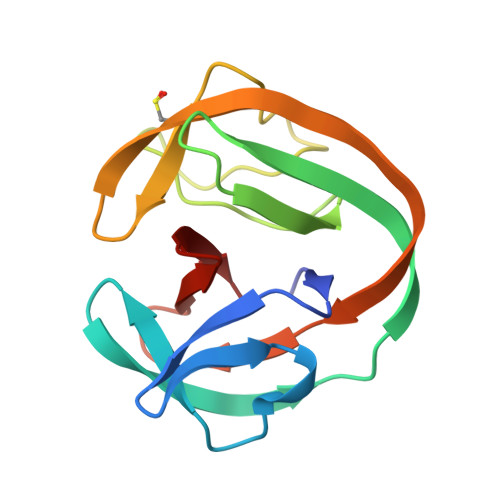
6RIZ

Crystal structure of gp41-1 intein (C1A, F65W, D107C)


Experimental Data Snapshot

  • Method: X-RAY DIFFRACTION
  • Resolution: 1.85 Å
  • R-Value Free: 0.202 
  • R-Value Work: 0.170 
  • R-Value Observed: 0.171 

wwPDB Validation   3D Report Full Report


This is version 1.2 of the entry. See complete history


Literature

The Convergence of the Hedgehog/Intein Fold in Different Protein Splicing Mechanisms.

Beyer, H.M.Virtanen, S.I.Aranko, A.S.Mikula, K.M.Lountos, G.T.Wlodawer, A.Ollila, O.H.S.Iwai, H.

(2020) Int J Mol Sci 21

  • DOI: https://doi.org/10.3390/ijms21218367
  • Primary Citation of Related Structures:  
    6RIX, 6RIY, 6RIZ

  • PubMed Abstract: 

    Protein splicing catalyzed by inteins utilizes many different combinations of amino-acid types at active sites. Inteins have been classified into three classes based on their characteristic sequences. We investigated the structural basis of the protein splicing mechanism of class 3 inteins by determining crystal structures of variants of a class 3 intein from Mycobacterium chimaera and molecular dynamics simulations, which suggested that the class 3 intein utilizes a different splicing mechanism from that of class 1 and 2 inteins. The class 3 intein uses a bond cleavage strategy reminiscent of proteases but share the same Hedgehog/INTein (HINT) fold of other intein classes. Engineering of class 3 inteins from a class 1 intein indicated that a class 3 intein would unlikely evolve directly from a class 1 or 2 intein. The HINT fold appears as structural and functional solution for trans -peptidyl and trans -esterification reactions commonly exploited by diverse mechanisms using different combinations of amino-acid types for the active-site residues.


  • Organizational Affiliation

    Institute of Biotechnology, University of Helsinki, P.O. Box 65, FIN-00014 Helsinki, Finland.


Macromolecules
Find similar proteins by:  (by identity cutoff)  |  3D Structure
Entity ID: 1
MoleculeChains Sequence LengthOrganismDetailsImage
gp41-1 intein128metagenomeMutation(s): 0 
Entity Groups  
Sequence Clusters30% Identity50% Identity70% Identity90% Identity95% Identity100% Identity
Sequence Annotations
Expand
  • Reference Sequence
Small Molecules
Modified Residues  1 Unique
IDChains TypeFormula2D DiagramParent
CSD
Query on CSD
A
L-PEPTIDE LINKINGC3 H7 N O4 SCYS
Experimental Data & Validation

Experimental Data

  • Method: X-RAY DIFFRACTION
  • Resolution: 1.85 Å
  • R-Value Free: 0.202 
  • R-Value Work: 0.170 
  • R-Value Observed: 0.171 
  • Space Group: P 65 2 2
Unit Cell:
Length ( Å )Angle ( ˚ )
a = 90.22α = 90
b = 90.22β = 90
c = 71.734γ = 120
Software Package:
Software NamePurpose
PHENIXrefinement
HKL-3000data reduction
HKL-3000data scaling
PHASERphasing
PHENIXmodel building

Structure Validation

View Full Validation Report


Entry History & Funding Information

Deposition Data


Funding OrganizationLocationGrant Number
Academy of FinlandFinland137995, 277335
Novo Nordisk FoundationFinlandNNF17OC0025402
Sigrid Juselius FoundationFinland--

Revision History  (Full details and data files)

  • Version 1.0: 2020-04-29
    Type: Initial release
  • Version 1.1: 2020-12-23
    Changes: Database references
  • Version 1.2: 2024-01-24
    Changes: Data collection, Database references, Refinement description