6RF8 | pdb_00006rf8

Cryo-EM structure of the N-terminal DC repeat (NDC) of NDC-NDC chimera (human sequence) bound to 13-protofilament GDP-microtubule


Experimental Data Snapshot

  • Method: ELECTRON MICROSCOPY
  • Resolution: 3.80 Å
  • Aggregation State: FILAMENT 
  • Reconstruction Method: SINGLE PARTICLE 

Starting Model: experimental
View more details

wwPDB Validation 3D Report Full Report

Validation slider image for 6RF8

This is version 1.1 of the entry. See complete history

Literature

A microtubule RELION-based pipeline for cryo-EM image processing.

Cook, A.D.Manka, S.W.Wang, S.Moores, C.A.Atherton, J.

(2020) J Struct Biol 209: 107402-107402

  • DOI: https://doi.org/10.1016/j.jsb.2019.10.004
  • Primary Citation Related Structures: 
    6RF8

  • PubMed Abstract: 

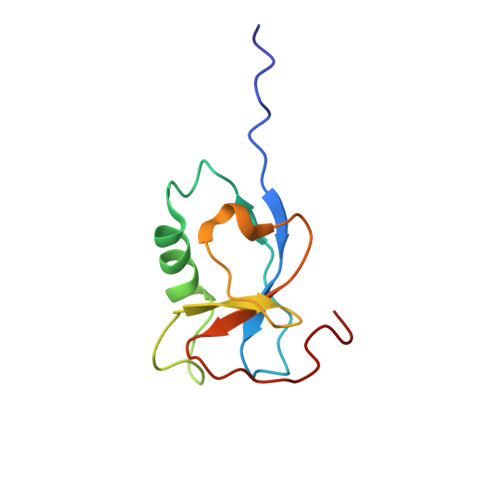
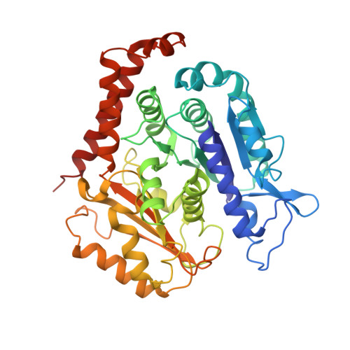
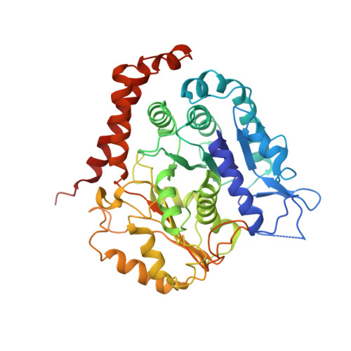
    Microtubules are polar filaments built from αβ-tubulin heterodimers that exhibit a range of architectures in vitro and in vivo. Tubulin heterodimers are arranged helically in the microtubule wall but many physiologically relevant architectures exhibit a break in helical symmetry known as the seam. Noisy 2D cryo-electron microscopy projection images of pseudo-helical microtubules therefore depict distinct but highly similar views owing to the high structural similarity of α- and β-tubulin. The determination of the αβ-tubulin register and seam location during image processing is essential for alignment accuracy that enables determination of biologically relevant structures. Here we present a pipeline designed for image processing and high-resolution reconstruction of cryo-electron microscopy microtubule datasets, based in the popular and user-friendly RELION image-processing package, Microtubule RELION-based Pipeline (MiRP). The pipeline uses a combination of supervised classification and prior knowledge about geometric lattice constraints in microtubules to accurately determine microtubule architecture and seam location. The presented method is fast and semi-automated, producing near-atomic resolution reconstructions with test datasets that contain a range of microtubule architectures and binding proteins.


  • Organizational Affiliation
    • Institute of Structural and Molecular Biology, Birkbeck, University of London, Malet Street, London, United Kingdom.

Macromolecule Content 

  • Total Structure Weight: 206.18 kDa 
  • Atom Count: 14,407 
  • Modeled Residue Count: 1,821 
  • Deposited Residue Count: 1,821 
  • Unique protein chains: 3

Macromolecules

Find similar proteins by:|  3D Structure
Entity ID: 1
MoleculeChains  Sequence LengthOrganismDetailsImage
Neuronal migration protein doublecortinA [auth N]99Homo sapiensMutation(s): 0 
Gene Names: DCXDBCNLISX
UniProt & NIH Common Fund Data Resources
Find proteins for O43602 (Homo sapiens)
Explore O43602 
Go to UniProtKB:  O43602
PHAROS:  O43602
GTEx:  ENSG00000077279 
Entity Groups
Sequence Clusters30% Identity50% Identity70% Identity90% Identity95% Identity100% Identity
UniProt GroupO43602
Sequence Annotations
Expand
Reference Sequence
Find similar proteins by:|  3D Structure
Entity ID: 2
MoleculeChains  Sequence LengthOrganismDetailsImage
Tubulin beta-2B chainB [auth b],
D [auth B]
429Bos taurusMutation(s): 0 
UniProt
Find proteins for Q6B856 (Bos taurus)
Explore Q6B856 
Go to UniProtKB:  Q6B856
Entity Groups
Sequence Clusters30% Identity50% Identity70% Identity90% Identity95% Identity100% Identity
UniProt GroupQ6B856
Sequence Annotations
Expand
Reference Sequence
Find similar proteins by:|  3D Structure
Entity ID: 3
MoleculeChains  Sequence LengthOrganismDetailsImage
Tubulin alpha-1B chainC [auth A],
E [auth a]
432Bos taurusMutation(s): 0 
EC: 3.6.5
UniProt
Find proteins for P81947 (Bos taurus)
Explore P81947 
Go to UniProtKB:  P81947
Entity Groups
Sequence Clusters30% Identity50% Identity70% Identity90% Identity95% Identity100% Identity
UniProt GroupP81947
Sequence Annotations
Expand
Reference Sequence

Experimental Data & Validation

Experimental Data

  • Method: ELECTRON MICROSCOPY
  • Resolution: 3.80 Å
  • Aggregation State: FILAMENT 
  • Reconstruction Method: SINGLE PARTICLE 

Structure Validation

View Full Validation Report


Entry History 

& Funding Information

Deposition Data

  • Released Date: 2020-04-15 
  • Deposition Author(s): Manka, S.W.

Funding OrganizationLocationGrant Number
Medical Research Council (United Kingdom)United KingdomMR/R000352/1

Revision History  (Full details and data files)

  • Version 1.0: 2020-04-15
    Type: Initial release
  • Version 1.1: 2024-05-22
    Changes: Data collection, Database references, Refinement description