6REK | pdb_00006rek

Crystal structure of Pizza6-SH with Cu2+


Experimental Data Snapshot

  • Method: X-RAY DIFFRACTION
  • Resolution: 1.10 Å
  • R-Value Free: 
    0.163 (Depositor), 0.169 (DCC) 
  • R-Value Work: 
    0.141 (Depositor) 
  • R-Value Observed: 
    0.142 (Depositor) 

Starting Model: experimental
View more details

wwPDB Validation 3D Report Full Report

Validation slider image for 6REK

This is version 1.1 of the entry. See complete history

Literature

Artificial beta-propeller protein-based hydrolases.

Clarke, D.E.Noguchi, H.Gryspeerdt, J.A.G.De Feyter, S.Voet, A.R.D.

(2019) Chem Commun (Camb) 55: 8880-8883

  • DOI: https://doi.org/10.1039/c9cc04388h
  • Primary Citation Related Structures: 
    6REG, 6REH, 6REJ, 6REK, 6REN

  • PubMed Abstract: 

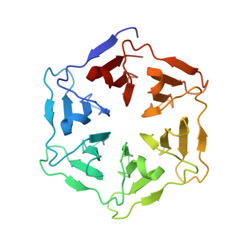
    We developed an artificial hydrolase based on the symmetrical Pizza6 β-propeller protein for the metal-free hydrolysis of 4-nitrophenyl acetate and butyrate. Through site-specific mutagenesis and crystallisation studies, the catalytic mechanism was investigated and found to be dependent on a threonine-histidine dyad. The mutant with additional histidine residues generated the highest kcat values, forming a His-His-Thr triad and matched previously reported metalloenzymes. The highly symmetrical β-propeller artificial enzymes and their protein-metal complexes have potential to be utilised in bioinorganic and supramolecular chemistry, as well as being developed further into 2D/3D catalytic materials.


  • Organizational Affiliation
    • Division of Molecular Imaging and Photonics, Department of Chemistry, KU Leuven, Celestijnenlaan 200F, 3001, Leuven, Belgium.

Macromolecule Content 

  • Total Structure Weight: 26.16 kDa 
  • Atom Count: 2,284 
  • Modeled Residue Count: 251 
  • Deposited Residue Count: 256 
  • Unique protein chains: 1

Macromolecules

Find similar proteins by:|  3D Structure
Entity ID: 1
MoleculeChains  Sequence LengthOrganismDetailsImage
Pizza6-SH256synthetic constructMutation(s): 0 

Experimental Data & Validation

Experimental Data

  • Method: X-RAY DIFFRACTION
  • Resolution: 1.10 Å
  • R-Value Free:  0.163 (Depositor), 0.169 (DCC) 
  • R-Value Work:  0.141 (Depositor) 
  • R-Value Observed: 0.142 (Depositor) 
Space Group: P 1 21 1
Unit Cell:
Length ( Å )Angle ( ˚ )
a = 32.708α = 90
b = 69.547β = 109.16
c = 48.266γ = 90
Software Package:
Software NamePurpose
PHENIXrefinement
XDSdata reduction
Aimlessdata scaling
PHASERphasing

Structure Validation

View Full Validation Report


Entry History 

& Funding Information

Deposition Data


Funding OrganizationLocationGrant Number
Research Foundation - FlandersBelgiumG0E4717N, G0F9316N, G051917N

Revision History  (Full details and data files)

  • Version 1.0: 2019-07-31
    Type: Initial release
  • Version 1.1: 2024-01-24
    Changes: Data collection, Database references, Derived calculations, Refinement description