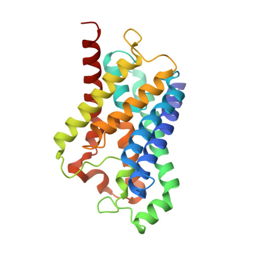
6QZJ

Crystal structure of human Aquaporin 7 at 2.2 A resolution


Experimental Data Snapshot

  • Method: X-RAY DIFFRACTION
  • Resolution: 2.20 Å
  • R-Value Free: 0.243 
  • R-Value Work: 0.209 
  • R-Value Observed: 0.210 

Starting Model: experimental
View more details

wwPDB Validation   3D Report Full Report


Ligand Structure Quality Assessment 


This is version 1.3 of the entry. See complete history


Literature

Structural Basis for Glycerol Efflux and Selectivity of Human Aquaporin 7.

de Mare, S.W.Venskutonyte, R.Eltschkner, S.de Groot, B.L.Lindkvist-Petersson, K.

(2020) Structure 28: 215-222.e3

  • DOI: https://doi.org/10.1016/j.str.2019.11.011
  • Primary Citation of Related Structures:  
    6QZI, 6QZJ

  • PubMed Abstract: 

    The aquaglyceroporin 7 (AQP7) facilitates permeation of glycerol through cell membranes and is crucial for lipid metabolism in humans. Glycerol efflux in human adipocytes is controlled by translocation of AQP7 to the plasma membrane upon hormone stimulation. Here we present two X-ray structures of human AQP7 at 1.9 and 2.2 Å resolution. The structures combined with molecular dynamics simulations suggest that AQP7 is a channel selective for glycerol and that glycerol may hamper water permeation through the channel. Moreover, the high resolution of the structures facilitated a detailed analysis of the orientation of glycerol in the pore, disclosing unusual positions of the hydroxyl groups. The data suggest that glycerol is conducted by a partly rotating movement through the channel. These observations provide a framework for understanding the basis of glycerol efflux and selectivity in aquaglyceroporins and pave the way for future design of AQP7 inhibitors.


  • Organizational Affiliation

    Experimental Medical Science, Medical Structural Biology, BMC C13, Lund University, SE-221 84 Lund, Sweden.


Macromolecules
Find similar proteins by:  (by identity cutoff)  |  3D Structure
Entity ID: 1
MoleculeChains Sequence LengthOrganismDetailsImage
Aquaporin-7247Homo sapiensMutation(s): 0 
Gene Names: AQP7AQP7LAQP9
Membrane Entity: Yes 
UniProt & NIH Common Fund Data Resources
Find proteins for O14520 (Homo sapiens)
Explore O14520 
Go to UniProtKB:  O14520
PHAROS:  O14520
GTEx:  ENSG00000165269 
Entity Groups  
Sequence Clusters30% Identity50% Identity70% Identity90% Identity95% Identity100% Identity
UniProt GroupO14520
Sequence Annotations
Expand
  • Reference Sequence
Small Molecules
Ligands 2 Unique
IDChains Name / Formula / InChI Key2D Diagram3D Interactions
PO4
Query on PO4

Download Ideal Coordinates CCD File 
M [auth A],
N [auth A]
PHOSPHATE ION
O4 P
NBIIXXVUZAFLBC-UHFFFAOYSA-K
GOL (Subject of Investigation/LOI)
Query on GOL

Download Ideal Coordinates CCD File 
B [auth A]
C [auth A]
D [auth A]
E [auth A]
F [auth A]
B [auth A],
C [auth A],
D [auth A],
E [auth A],
F [auth A],
G [auth A],
H [auth A],
I [auth A],
J [auth A],
K [auth A],
L [auth A]
GLYCEROL
C3 H8 O3
PEDCQBHIVMGVHV-UHFFFAOYSA-N
Experimental Data & Validation

Experimental Data

  • Method: X-RAY DIFFRACTION
  • Resolution: 2.20 Å
  • R-Value Free: 0.243 
  • R-Value Work: 0.209 
  • R-Value Observed: 0.210 
  • Space Group: I 4
Unit Cell:
Length ( Å )Angle ( ˚ )
a = 86.761α = 90
b = 86.761β = 90
c = 83.544γ = 90
Software Package:
Software NamePurpose
XDSdata reduction
Aimlessdata scaling
PHASERphasing
PHENIXrefinement
PDB_EXTRACTdata extraction

Structure Validation

View Full Validation Report


Ligand Structure Quality Assessment 


Entry History & Funding Information

Deposition Data


Funding OrganizationLocationGrant Number
Swedish Research CouncilSweden--

Revision History  (Full details and data files)

  • Version 1.0: 2019-11-27
    Type: Initial release
  • Version 1.1: 2019-12-25
    Changes: Database references
  • Version 1.2: 2020-02-12
    Changes: Database references
  • Version 1.3: 2024-01-24
    Changes: Data collection, Database references, Refinement description