6QVA

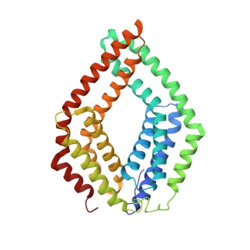
Crystal structure of a CHAD domain from Chlorobium tepidum in complex with inorganic polyphosphate


Experimental Data Snapshot

  • Method: X-RAY DIFFRACTION
  • Resolution: 2.05 Å
  • R-Value Free: 0.255 
  • R-Value Work: 0.208 
  • R-Value Observed: 0.210 

wwPDB Validation   3D Report Full Report


Ligand Structure Quality Assessment 


This is version 1.2 of the entry. See complete history


Literature

Molecular characterization of CHAD domains as inorganic polyphosphate-binding modules.

Lorenzo-Orts, L.Hohmann, U.Zhu, J.Hothorn, M.

(2019) Life Sci Alliance 2

  • DOI: https://doi.org/10.26508/lsa.201900385
  • Primary Citation of Related Structures:  
    6QV5, 6QV7, 6QVA

  • PubMed Abstract: 

    Inorganic polyphosphates (polyPs) are linear polymers of orthophosphate units linked by phosphoanhydride bonds. Here, we report that bacterial, archaeal, and eukaryotic conserved histidine α-helical (CHAD) domains are specific polyP-binding modules. Crystal structures reveal that CHAD domains are formed by two four-helix bundles, giving rise to a central pore surrounded by conserved basic surface patches. Different CHAD domains bind polyPs with dissociation constants ranging from the nano- to mid-micromolar range, but not nucleic acids. A CHAD-polyP complex structure reveals the phosphate polymer binding across the central pore and along the two basic patches. Mutational analysis of CHAD-polyP interface residues validates the complex structure. The presence of a CHAD domain in the polyPase ygiF enhances its enzymatic activity. The only known CHAD protein from the plant Ricinus communis localizes to the nucleus/nucleolus when expressed in Arabidopsis and tobacco, suggesting that plants may harbor polyPs in these compartments. We propose that CHAD domains may be used to engineer the properties of polyP-metabolizing enzymes and to specifically localize polyP stores in eukaryotic cells and tissues.


  • Organizational Affiliation

    Structural Plant Biology Laboratory, Department of Botany and Plant Biology, Faculty of Sciences, University of Geneva, Geneva, Switzerland.


Macromolecules
Find similar proteins by:  (by identity cutoff)  |  3D Structure
Entity ID: 1
MoleculeChains Sequence LengthOrganismDetailsImage
CHAD domain318Chlorobaculum tepidum TLSMutation(s): 0 
Gene Names: CT0884
UniProt
Find proteins for Q8KE09 (Chlorobaculum tepidum (strain ATCC 49652 / DSM 12025 / NBRC 103806 / TLS))
Explore Q8KE09 
Go to UniProtKB:  Q8KE09
Entity Groups  
Sequence Clusters30% Identity50% Identity70% Identity90% Identity95% Identity100% Identity
UniProt GroupQ8KE09
Sequence Annotations
Expand
  • Reference Sequence
Small Molecules
Ligands 4 Unique
IDChains Name / Formula / InChI Key2D Diagram3D Interactions
9PI
Query on 9PI

Download Ideal Coordinates CCD File 
F [auth A]bis[oxidanyl-[oxidanyl-[oxidanyl(phosphonooxy)phosphoryl]oxy-phosphoryl]oxy-phosphoryl] hydrogen phosphate
H11 O28 P9
CGJFWWJXTCZYEB-UHFFFAOYSA-N
3PO
Query on 3PO

Download Ideal Coordinates CCD File 
G [auth A]TRIPHOSPHATE
H5 O10 P3
UNXRWKVEANCORM-UHFFFAOYSA-N
PO4
Query on PO4

Download Ideal Coordinates CCD File 
H [auth A]PHOSPHATE ION
O4 P
NBIIXXVUZAFLBC-UHFFFAOYSA-K
EDO
Query on EDO

Download Ideal Coordinates CCD File 
B [auth A],
C [auth A],
D [auth A],
E [auth A]
1,2-ETHANEDIOL
C2 H6 O2
LYCAIKOWRPUZTN-UHFFFAOYSA-N
Binding Affinity Annotations 
IDSourceBinding Affinity
9PI Binding MOAD:  6QVA IC50: 1000 (nM) from 1 assay(s)
Experimental Data & Validation

Experimental Data

  • Method: X-RAY DIFFRACTION
  • Resolution: 2.05 Å
  • R-Value Free: 0.255 
  • R-Value Work: 0.208 
  • R-Value Observed: 0.210 
  • Space Group: I 2 2 2
Unit Cell:
Length ( Å )Angle ( ˚ )
a = 69.4α = 90
b = 97.031β = 90
c = 106.397γ = 90
Software Package:
Software NamePurpose
REFMACrefinement
XDSdata reduction
XDSdata scaling
PHASERphasing

Structure Validation

View Full Validation Report


Ligand Structure Quality Assessment 


Entry History & Funding Information

Deposition Data


Funding OrganizationLocationGrant Number
European Research CouncilSwitzerland310856

Revision History  (Full details and data files)

  • Version 1.0: 2019-05-29
    Type: Initial release
  • Version 1.1: 2019-06-05
    Changes: Data collection, Database references
  • Version 1.2: 2024-01-24
    Changes: Data collection, Database references, Refinement description