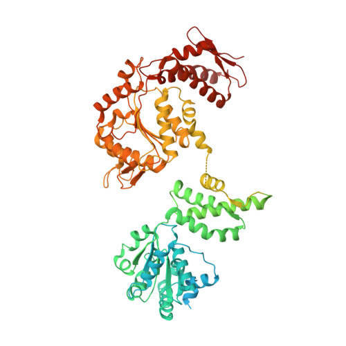
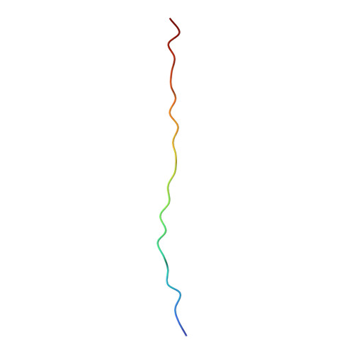
6QS8

ClpB (DWB and K476C mutant) bound to casein in presence of ATPgammaS - state KC-2B


Experimental Data Snapshot

  • Method: ELECTRON MICROSCOPY
  • Resolution: 3.90 Å
  • Aggregation State: PARTICLE 
  • Reconstruction Method: SINGLE PARTICLE 

Starting Model: experimental
View more details

wwPDB Validation   3D Report Full Report


This is version 1.5 of the entry. See complete history


Literature

Two-Step Activation Mechanism of the ClpB Disaggregase for Sequential Substrate Threading by the Main ATPase Motor.

Deville, C.Franke, K.Mogk, A.Bukau, B.Saibil, H.R.

(2019) Cell Rep 27: 3433-3446.e4

  • DOI: https://doi.org/10.1016/j.celrep.2019.05.075
  • Primary Citation of Related Structures:  
    6QS4, 6QS6, 6QS7, 6QS8, 6RN2, 6RN3, 6RN4

  • PubMed Abstract: 

    AAA+ proteins form asymmetric hexameric rings that hydrolyze ATP and thread substrate proteins through a central channel via mobile substrate-binding pore loops. Understanding how ATPase and threading activities are regulated and intertwined is key to understanding the AAA+ protein mechanism. We studied the disaggregase ClpB, which contains tandem ATPase domains (AAA1, AAA2) and shifts between low and high ATPase and threading activities. Coiled-coil M-domains repress ClpB activity by encircling the AAA1 ring. Here, we determine the mechanism of ClpB activation by comparing ATPase mechanisms and cryo-EM structures of ClpB wild-type and a constitutively active ClpB M-domain mutant. We show that ClpB activation reduces ATPase cooperativity and induces a sequential mode of ATP hydrolysis in the AAA2 ring, the main ATPase motor. AAA1 and AAA2 rings do not work synchronously but in alternating cycles. This ensures high grip, enabling substrate threading via a processive, rope-climbing mechanism.


  • Organizational Affiliation

    Department of Crystallography, Institute of Structural and Molecular Biology, Birkbeck, University of London, Malet Street, London WC1E 7HX, UK.


Macromolecules
Find similar proteins by:  (by identity cutoff)  |  3D Structure
Entity ID: 1
MoleculeChains Sequence LengthOrganismDetailsImage
Chaperone protein ClpB
A, B, C, D, E
A, B, C, D, E, F
858Escherichia coliMutation(s): 3 
Gene Names: clpBhtpMb2592JW2573
UniProt
Find proteins for P63284 (Escherichia coli (strain K12))
Explore P63284 
Go to UniProtKB:  P63284
Entity Groups  
Sequence Clusters30% Identity50% Identity70% Identity90% Identity95% Identity100% Identity
UniProt GroupP63284
Sequence Annotations
Expand
  • Reference Sequence

Find similar proteins by:  Sequence   |   3D Structure  

Entity ID: 2
MoleculeChains Sequence LengthOrganismDetailsImage
caseinG [auth S]24Bos taurusMutation(s): 0 
Sequence Annotations
Expand
  • Reference Sequence
Small Molecules
Ligands 3 Unique
IDChains Name / Formula / InChI Key2D Diagram3D Interactions
AGS
Query on AGS

Download Ideal Coordinates CCD File 
H [auth A]
J [auth B]
K [auth B]
N [auth C]
O [auth C]
H [auth A],
J [auth B],
K [auth B],
N [auth C],
O [auth C],
R [auth D],
S [auth D],
V [auth E],
W [auth E],
Z [auth F]
PHOSPHOTHIOPHOSPHORIC ACID-ADENYLATE ESTER
C10 H16 N5 O12 P3 S
NLTUCYMLOPLUHL-KQYNXXCUSA-N
ADP
Query on ADP

Download Ideal Coordinates CCD File 
Y [auth F]ADENOSINE-5'-DIPHOSPHATE
C10 H15 N5 O10 P2
XTWYTFMLZFPYCI-KQYNXXCUSA-N
MG
Query on MG

Download Ideal Coordinates CCD File 
I [auth A]
L [auth B]
M [auth B]
P [auth C]
Q [auth C]
I [auth A],
L [auth B],
M [auth B],
P [auth C],
Q [auth C],
T [auth D],
U [auth D],
X [auth E]
MAGNESIUM ION
Mg
JLVVSXFLKOJNIY-UHFFFAOYSA-N
Experimental Data & Validation

Experimental Data

  • Method: ELECTRON MICROSCOPY
  • Resolution: 3.90 Å
  • Aggregation State: PARTICLE 
  • Reconstruction Method: SINGLE PARTICLE 
EM Software:
TaskSoftware PackageVersion
MODEL REFINEMENTPHENIX1.12
RECONSTRUCTIONRELION2.1

Structure Validation

View Full Validation Report


Entry History & Funding Information

Deposition Data


Funding OrganizationLocationGrant Number
Wellcome TrustUnited Kingdom106252
Wellcome TrustUnited Kingdom101488
Wellcome TrustUnited Kingdom079605

Revision History  (Full details and data files)

  • Version 1.0: 2019-07-03
    Type: Initial release
  • Version 1.1: 2019-07-10
    Changes: Data collection
  • Version 1.2: 2019-07-17
    Changes: Data collection, Data processing
  • Version 1.3: 2019-08-21
    Changes: Data collection, Experimental preparation
  • Version 1.4: 2019-12-18
    Changes: Other
  • Version 1.5: 2024-05-15
    Changes: Data collection, Database references, Refinement description