6QMU

A tetrahedral boronic acid diester formed by a non-natural amino acid in the ligand pocket of an engineered lipocalin


Experimental Data Snapshot

  • Method: X-RAY DIFFRACTION
  • Resolution: 1.98 Å
  • R-Value Free: 0.271 
  • R-Value Work: 0.207 
  • R-Value Observed: 0.210 

wwPDB Validation   3D Report Full Report


This is version 1.3 of the entry. See complete history


Literature

A Tetrahedral Boronic Acid Diester Formed by an Unnatural Amino Acid in the Ligand Pocket of an Engineered Lipocalin.

Sommer, C.A.Eichinger, A.Skerra, A.

(2020) Chembiochem 21: 469-472

  • DOI: https://doi.org/10.1002/cbic.201900405
  • Primary Citation of Related Structures:  
    6QMU

  • PubMed Abstract: 

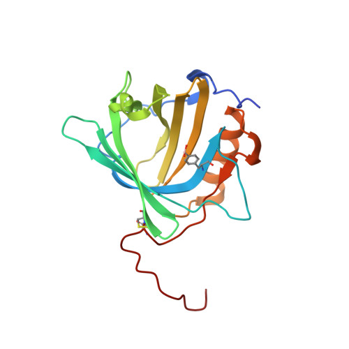
    Boronic acids have long been known to form cyclic diesters with cis-diol compounds, including many carbohydrates. This phenomenon was previously exploited to create an artificial lectin by incorporating p-borono-l-phenylalanine (Bpa) into the ligand pocket of an engineered lipocalin, resulting in a so-called Borocalin. Here we describe the X-ray analysis of its covalent complex with 4-nitrocatechol as a high-affinity model ligand. As expected, the crystal structure reveals the formation of a cyclic diester between the biosynthetic boronate side chain and the two ortho-hydroxy substituents of the benzene ring. Interestingly, the boron also has a hydroxide ion associated, despite an only moderately basic pH 8.5 in the crystallization buffer. The complex is stabilized by a polar contact to the side chain of Asn134 within the ligand pocket, thus validating the functional design of the Borocalin as an artificial sugar-binding protein. Our structural analysis demonstrates how a boronate can form a thermodynamically stable diester with a vicinal diol in a tetrahedral configuration in aqueous solution near physiological pH. Moreover, our data provide a basis for the further engineering of the Borocalin with the goal of specific recognition of biologically relevant glycans.


  • Organizational Affiliation

    Lehrstuhl für Biologische Chemie, Technische Universität München, Emil-Erlenmeyer-Forum 5, 85354, Freising, Germany.


Macromolecules
Find similar proteins by:  (by identity cutoff)  |  3D Structure
Entity ID: 1
MoleculeChains Sequence LengthOrganismDetailsImage
Neutrophil gelatinase-associated lipocalin
A, B
188Homo sapiensMutation(s): 6 
Gene Names: LCN2HNLNGAL
UniProt & NIH Common Fund Data Resources
Find proteins for P80188 (Homo sapiens)
Explore P80188 
Go to UniProtKB:  P80188
PHAROS:  P80188
GTEx:  ENSG00000148346 
Entity Groups  
Sequence Clusters30% Identity50% Identity70% Identity90% Identity95% Identity100% Identity
UniProt GroupP80188
Sequence Annotations
Expand
  • Reference Sequence
Small Molecules
Modified Residues  1 Unique
IDChains TypeFormula2D DiagramParent
7N8
Query on 7N8
A, B
L-PEPTIDE LINKINGC9 H12 B N O4PHE
Experimental Data & Validation

Experimental Data

  • Method: X-RAY DIFFRACTION
  • Resolution: 1.98 Å
  • R-Value Free: 0.271 
  • R-Value Work: 0.207 
  • R-Value Observed: 0.210 
  • Space Group: C 1 2 1
Unit Cell:
Length ( Å )Angle ( ˚ )
a = 130.486α = 90
b = 44.15β = 133.47
c = 92.167γ = 90
Software Package:
Software NamePurpose
MOSFLMdata reduction
SCALAdata scaling
PHASERphasing
REFMACrefinement
PDB_EXTRACTdata extraction

Structure Validation

View Full Validation Report


Entry History & Funding Information

Deposition Data


Funding OrganizationLocationGrant Number
German Research FoundationGermanyGRK2062

Revision History  (Full details and data files)

  • Version 1.0: 2019-08-28
    Type: Initial release
  • Version 1.1: 2019-11-20
    Changes: Database references
  • Version 1.2: 2020-03-04
    Changes: Database references
  • Version 1.3: 2024-01-24
    Changes: Data collection, Database references, Derived calculations, Refinement description