6QGS

Crystal structure of APT1 bound to palmitic acid.


Experimental Data Snapshot

  • Method: X-RAY DIFFRACTION
  • Resolution: 2.75 Å
  • R-Value Free: 0.230 
  • R-Value Work: 0.184 
  • R-Value Observed: 0.186 

wwPDB Validation   3D Report Full Report


Ligand Structure Quality Assessment 


This is version 1.3 of the entry. See complete history


Literature

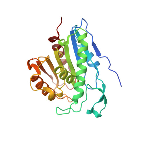
Palmitoylated acyl protein thioesterase APT2 deforms membranes to extract substrate acyl chains.

Abrami, L.Audagnotto, M.Ho, S.Marcaida, M.J.Mesquita, F.S.Anwar, M.U.Sandoz, P.A.Fonti, G.Pojer, F.Dal Peraro, M.van der Goot, F.G.

(2021) Nat Chem Biol 

  • DOI: https://doi.org/10.1038/s41589-021-00753-2
  • Primary Citation of Related Structures:  
    6QGN, 6QGO, 6QGQ, 6QGS

  • PubMed Abstract: 

    Many biochemical reactions require controlled recruitment of proteins to membranes. This is largely regulated by posttranslational modifications. A frequent one is S-acylation, which consists of the addition of acyl chains and can be reversed by poorly understood acyl protein thioesterases (APTs). Using a panel of computational and experimental approaches, we dissect the mode of action of the major cellular thioesterase APT2 (LYPLA2). We show that soluble APT2 is vulnerable to proteasomal degradation, from which membrane binding protects it. Interaction with membranes requires three consecutive steps: electrostatic attraction, insertion of a hydrophobic loop and S-acylation by the palmitoyltransferases ZDHHC3 or ZDHHC7. Once bound, APT2 is predicted to deform the lipid bilayer to extract the acyl chain bound to its substrate and capture it in a hydrophobic pocket to allow hydrolysis. This molecular understanding of APT2 paves the way to understand the dynamics of APT2-mediated deacylation of substrates throughout the endomembrane system.


  • Organizational Affiliation

    Global Health Institute, School of Life Sciences, EPFL, Lausanne, Switzerland.


Macromolecules
Find similar proteins by:  (by identity cutoff)  |  3D Structure
Entity ID: 1
MoleculeChains Sequence LengthOrganismDetailsImage
Acyl-protein thioesterase 1233Homo sapiensMutation(s): 0 
Gene Names: LYPLA1APT1LPL1
EC: 3.1.2
Membrane Entity: Yes 
UniProt & NIH Common Fund Data Resources
Find proteins for O75608 (Homo sapiens)
Explore O75608 
Go to UniProtKB:  O75608
PHAROS:  O75608
GTEx:  ENSG00000120992 
Entity Groups  
Sequence Clusters30% Identity50% Identity70% Identity90% Identity95% Identity100% Identity
UniProt GroupO75608
Sequence Annotations
Expand
  • Reference Sequence
Experimental Data & Validation

Experimental Data

  • Method: X-RAY DIFFRACTION
  • Resolution: 2.75 Å
  • R-Value Free: 0.230 
  • R-Value Work: 0.184 
  • R-Value Observed: 0.186 
  • Space Group: P 41 21 2
Unit Cell:
Length ( Å )Angle ( ˚ )
a = 81.66α = 90
b = 81.66β = 90
c = 441.946γ = 90
Software Package:
Software NamePurpose
PHENIXrefinement
XDSdata reduction
XDSdata scaling
PHASERphasing

Structure Validation

View Full Validation Report


Ligand Structure Quality Assessment 


Entry History & Funding Information

Deposition Data


Funding OrganizationLocationGrant Number
Swiss National Science FoundationSwitzerland200020_157153

Revision History  (Full details and data files)

  • Version 1.0: 2020-02-05
    Type: Initial release
  • Version 1.1: 2021-03-17
    Changes: Database references
  • Version 1.2: 2021-03-31
    Changes: Database references
  • Version 1.3: 2024-01-24
    Changes: Data collection, Database references, Refinement description