6QF7

Crystal structures of the recombinant beta-Factor XIIa protease with bound Thr-Arg and Pro-Arg substrate mimetics


Experimental Data Snapshot

  • Method: X-RAY DIFFRACTION
  • Resolution: 4.00 Å
  • R-Value Free: 0.356 
  • R-Value Work: 0.297 
  • R-Value Observed: 0.303 

wwPDB Validation   3D Report Full Report


Ligand Structure Quality Assessment 


This is version 2.0 of the entry. See complete history


Literature

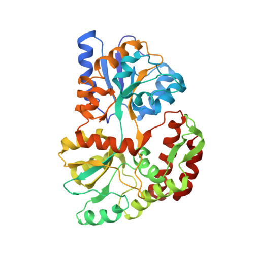
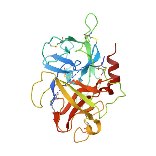
Crystal structures of the recombinant beta-factor XIIa protease with bound Thr-Arg and Pro-Arg substrate mimetics.

Pathak, M.Manna, R.Li, C.Kaira, B.G.Hamad, B.K.Belviso, B.D.Bonturi, C.R.Dreveny, I.Fischer, P.M.Dekker, L.V.Oliva, M.L.V.Emsley, J.

(2019) Acta Crystallogr D Struct Biol 75: 578-591

  • DOI: https://doi.org/10.1107/S2059798319006910
  • Primary Citation of Related Structures:  
    6GT6, 6QF7

  • PubMed Abstract: 

    Coagulation factor XII (FXII) is a key initiator of the contact pathway, which contributes to inflammatory pathways. FXII circulates as a zymogen, which when auto-activated forms factor XIIa (FXIIa). Here, the production of the recombinant FXIIa protease domain (βFXIIa His ) with yields of ∼1-2 mg per litre of insect-cell culture is reported. A second construct utilized an N-terminal maltose-binding protein (MBP) fusion (MBP-βFXIIa His ). Crystal structures were determined of MBP-βFXIIa His in complex with the inhibitor D-Phe-Pro-Arg chloromethyl ketone (PPACK) and of βFXIIa His in isolation. The βFXIIa His structure revealed that the S2 and S1 pockets were occupied by Thr and Arg residues, respectively, from an adjacent molecule in the crystal. The Thr-Arg sequence mimics the P2-P1 FXIIa cleavage-site residues present in the natural substrates prekallikrein and FXII, and Pro-Arg (from PPACK) mimics the factor XI cleavage site. A comparison of the βFXIIa His structure with the available crystal structure of the zymogen-like FXII protease revealed large conformational changes centred around the S1 pocket and an alternate conformation for the 99-loop, Tyr99 and the S2 pocket. Further comparison with activated protease structures of factors IXa and Xa, which also have the Tyr99 residue, reveals that a more open form of the S2 pocket only occurs in the presence of a substrate mimetic. The FXIIa inhibitors EcTI and infestin-4 have Pro-Arg and Phe-Arg P2-P1 sequences, respectively, and the interactions that these inhibitors make with βFXIIa are also described. These structural studies of βFXIIa provide insight into substrate and inhibitor recognition and establish a scaffold for the structure-guided drug design of novel antithrombotic and anti-inflammatory agents.


  • Organizational Affiliation

    Centre for Biomolecular Sciences, School of Pharmacy, University of Nottingham, Nottingham NG7 2RD, England.


Macromolecules
Find similar proteins by:  (by identity cutoff)  |  3D Structure
Entity ID: 1
MoleculeChains Sequence LengthOrganismDetailsImage
Maltodextrin-binding protein
A, C
374Escherichia coliMutation(s): 0 
Gene Names: malENCTC8450_00456NCTC9775_03059
Entity Groups  
Sequence Clusters30% Identity50% Identity70% Identity90% Identity95% Identity100% Identity
Sequence Annotations
Expand
  • Reference Sequence
Find similar proteins by:  (by identity cutoff)  |  3D Structure
Entity ID: 2
MoleculeChains Sequence LengthOrganismDetailsImage
Coagulation factor XII
B, D
254Homo sapiensMutation(s): 0 
Gene Names: F12
EC: 3.4.21.38
UniProt & NIH Common Fund Data Resources
Find proteins for P00748 (Homo sapiens)
Explore P00748 
Go to UniProtKB:  P00748
PHAROS:  P00748
GTEx:  ENSG00000131187 
Entity Groups  
Sequence Clusters30% Identity50% Identity70% Identity90% Identity95% Identity100% Identity
UniProt GroupP00748
Sequence Annotations
Expand
  • Reference Sequence
Oligosaccharides

Help

Entity ID: 3
MoleculeChains Length2D Diagram Glycosylation3D Interactions
2-acetamido-2-deoxy-beta-D-glucopyranose-(1-4)-2-acetamido-2-deoxy-beta-D-glucopyranose
E
2N/A
Glycosylation Resources
GlyTouCan:  G42666HT
GlyCosmos:  G42666HT
GlyGen:  G42666HT
Entity ID: 4
MoleculeChains Length2D Diagram Glycosylation3D Interactions
alpha-D-glucopyranose-(1-4)-alpha-D-glucopyranose
F, G
2N/A
Glycosylation Resources
GlyTouCan:  G07411ON
GlyCosmos:  G07411ON
Small Molecules
Ligands 2 Unique
IDChains Name / Formula / InChI Key2D Diagram3D Interactions
0G6
Query on 0G6

Download Ideal Coordinates CCD File 
I [auth B],
J [auth D]
D-phenylalanyl-N-[(2S,3S)-6-{[amino(iminio)methyl]amino}-1-chloro-2-hydroxyhexan-3-yl]-L-prolinamide
C21 H34 Cl N6 O3
DVFLYEYCMMLBTQ-VSZNYVQBSA-O
NAG
Query on NAG

Download Ideal Coordinates CCD File 
H [auth B]2-acetamido-2-deoxy-beta-D-glucopyranose
C8 H15 N O6
OVRNDRQMDRJTHS-FMDGEEDCSA-N
Experimental Data & Validation

Experimental Data

  • Method: X-RAY DIFFRACTION
  • Resolution: 4.00 Å
  • R-Value Free: 0.356 
  • R-Value Work: 0.297 
  • R-Value Observed: 0.303 
  • Space Group: P 41 21 2
Unit Cell:
Length ( Å )Angle ( ˚ )
a = 131.45α = 90
b = 131.45β = 90
c = 238.35γ = 90
Software Package:
Software NamePurpose
REFMACrefinement
XDSdata reduction
Aimlessdata scaling
PHASERphasing

Structure Validation

View Full Validation Report


Ligand Structure Quality Assessment 


Entry History & Funding Information

Deposition Data


Funding OrganizationLocationGrant Number
British Heart FoundationUnited KingdomRG/12/9/29775

Revision History  (Full details and data files)

  • Version 1.0: 2019-06-05
    Type: Initial release
  • Version 1.1: 2019-06-26
    Changes: Data collection, Database references
  • Version 1.2: 2019-07-31
    Changes: Data collection, Structure summary
  • Version 2.0: 2020-07-29
    Type: Remediation
    Reason: Carbohydrate remediation
    Changes: Advisory, Atomic model, Data collection, Derived calculations, Non-polymer description, Structure summary