6QDY

The crystal structure of Sporosarcina pasteurii urease in complex with its substrate urea


Experimental Data Snapshot

  • Method: X-RAY DIFFRACTION
  • Resolution: 1.42 Å
  • R-Value Free: 0.119 
  • R-Value Work: 0.095 
  • R-Value Observed: 0.096 

wwPDB Validation   3D Report Full Report


This is version 1.1 of the entry. See complete history


Literature

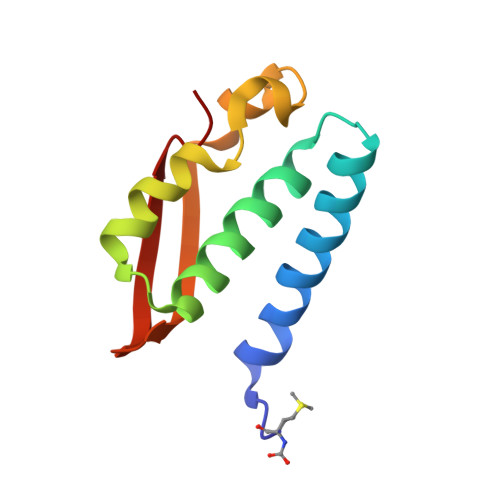
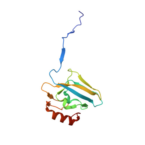
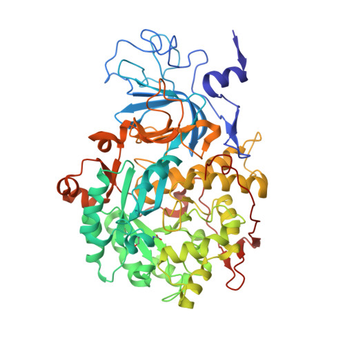
The Structure of the Elusive Urease-Urea Complex Unveils the Mechanism of a Paradigmatic Nickel-Dependent Enzyme.

Mazzei, L.Cianci, M.Benini, S.Ciurli, S.

(2019) Angew Chem Int Ed Engl 58: 7415-7419

  • DOI: https://doi.org/10.1002/anie.201903565
  • Primary Citation of Related Structures:  
    6QDY

  • PubMed Abstract: 

    Urease, the most efficient enzyme known, contains an essential dinuclear Ni II cluster in the active site. It catalyzes the hydrolysis of urea, inducing a rapid pH increase that has negative effects on human health and agriculture. Thus, the control of urease activity is of utmost importance in medical, pharmaceutical, and agro-environmental applications. All known urease inhibitors are either toxic or inefficient. The development of new and efficient chemicals able to inhibit urease relies on the knowledge of all steps of the catalytic mechanism. The short (microseconds) lifetime of the urease-urea complex has hampered the determination of its structure. The present study uses fluoride to substitute the hydroxide acting as the co-substrate in the reaction, preventing the occurrence of the catalytic steps that follow substrate binding. The 1.42 Å crystal structure of the urease-urea complex, reported here, resolves the enduring debate on the mechanism of this metalloenzyme.


  • Organizational Affiliation

    Laboratory of Bioinorganic Chemistry, Departement of Pharmacy and Biotechnology, University of Bologna, Via Giuseppe Fanin 40, 40138, Bologna, Italy.


Macromolecules
Find similar proteins by:  (by identity cutoff)  |  3D Structure
Entity ID: 1
MoleculeChains Sequence LengthOrganismDetailsImage
Urease subunit gamma100Sporosarcina pasteuriiMutation(s): 0 
EC: 3.5.1.5
UniProt
Find proteins for P41022 (Sporosarcina pasteurii)
Explore P41022 
Go to UniProtKB:  P41022
Entity Groups  
Sequence Clusters30% Identity50% Identity70% Identity90% Identity95% Identity100% Identity
UniProt GroupP41022
Sequence Annotations
Expand
  • Reference Sequence
Find similar proteins by:  (by identity cutoff)  |  3D Structure
Entity ID: 2
MoleculeChains Sequence LengthOrganismDetailsImage
Urease subunit beta122Sporosarcina pasteuriiMutation(s): 0 
EC: 3.5.1.5
UniProt
Find proteins for P41021 (Sporosarcina pasteurii)
Explore P41021 
Go to UniProtKB:  P41021
Entity Groups  
Sequence Clusters30% Identity50% Identity70% Identity90% Identity95% Identity100% Identity
UniProt GroupP41021
Sequence Annotations
Expand
  • Reference Sequence
Find similar proteins by:  (by identity cutoff)  |  3D Structure
Entity ID: 3
MoleculeChains Sequence LengthOrganismDetailsImage
Urease subunit alpha570Sporosarcina pasteuriiMutation(s): 0 
EC: 3.5.1.5
UniProt
Find proteins for P41020 (Sporosarcina pasteurii)
Explore P41020 
Go to UniProtKB:  P41020
Entity Groups  
Sequence Clusters30% Identity50% Identity70% Identity90% Identity95% Identity100% Identity
UniProt GroupP41020
Sequence Annotations
Expand
  • Reference Sequence
Small Molecules
Ligands 5 Unique
IDChains Name / Formula / InChI Key2D Diagram3D Interactions
SO4
Query on SO4

Download Ideal Coordinates CCD File 
I [auth B],
R [auth C]
SULFATE ION
O4 S
QAOWNCQODCNURD-UHFFFAOYSA-L
EDO
Query on EDO

Download Ideal Coordinates CCD File 
D [auth A]
E [auth A]
F [auth A]
G [auth B]
H [auth B]
D [auth A],
E [auth A],
F [auth A],
G [auth B],
H [auth B],
L [auth C],
M [auth C],
N [auth C],
O [auth C],
P [auth C],
Q [auth C]
1,2-ETHANEDIOL
C2 H6 O2
LYCAIKOWRPUZTN-UHFFFAOYSA-N
URE
Query on URE

Download Ideal Coordinates CCD File 
T [auth C]UREA
C H4 N2 O
XSQUKJJJFZCRTK-UHFFFAOYSA-N
NI
Query on NI

Download Ideal Coordinates CCD File 
J [auth C],
K [auth C]
NICKEL (II) ION
Ni
VEQPNABPJHWNSG-UHFFFAOYSA-N
F
Query on F

Download Ideal Coordinates CCD File 
S [auth C]FLUORIDE ION
F
KRHYYFGTRYWZRS-UHFFFAOYSA-M
Modified Residues  2 Unique
IDChains TypeFormula2D DiagramParent
CXM
Query on CXM
A
L-PEPTIDE LINKINGC6 H11 N O4 SMET
KCX
Query on KCX
C
L-PEPTIDE LINKINGC7 H14 N2 O4LYS
Experimental Data & Validation

Experimental Data

  • Method: X-RAY DIFFRACTION
  • Resolution: 1.42 Å
  • R-Value Free: 0.119 
  • R-Value Work: 0.095 
  • R-Value Observed: 0.096 
  • Space Group: P 63 2 2
Unit Cell:
Length ( Å )Angle ( ˚ )
a = 131.565α = 90
b = 131.565β = 90
c = 188.683γ = 120
Software Package:
Software NamePurpose
PHENIXrefinement
REFMACrefinement
XDSdata reduction
Aimlessdata scaling

Structure Validation

View Full Validation Report


Entry History 

Deposition Data

Revision History  (Full details and data files)

  • Version 1.0: 2019-11-13
    Type: Initial release
  • Version 1.1: 2024-01-24
    Changes: Data collection, Database references, Derived calculations, Refinement description