6QAF

Crystal structure of human Arginase-1 at pH 9.0 in complex with CB-1158/INCB001158


Experimental Data Snapshot

  • Method: X-RAY DIFFRACTION
  • Resolution: 1.61 Å
  • R-Value Free: 0.177 
  • R-Value Work: 0.147 
  • R-Value Observed: 0.149 

wwPDB Validation   3D Report Full Report


Ligand Structure Quality Assessment 


This is version 1.2 of the entry. See complete history


Literature

Structural insights into human Arginase-1 pH dependence and its inhibition by the small molecule inhibitor CB-1158.

Grobben, Y.Uitdehaag, J.C.M.Willemsen-Seegers, N.Tabak, W.W.A.de Man, J.Buijsman, R.C.Zaman, G.J.R.

(2020) J Struct Biol X 4: 100014-100014

  • DOI: https://doi.org/10.1016/j.yjsbx.2019.100014
  • Primary Citation of Related Structures:  
    6Q92, 6Q9P, 6QAF

  • PubMed Abstract: 

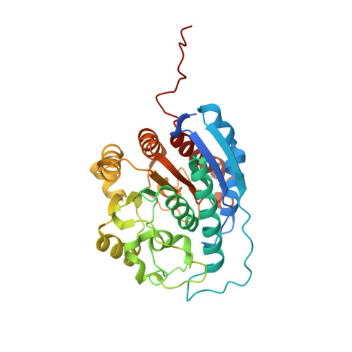
    Arginase-1 is a manganese-dependent metalloenzyme that catalyzes the hydrolysis of L-arginine into L-ornithine and urea. Arginase-1 is abundantly expressed by tumor-infiltrating myeloid cells that promote tumor immunosuppression, which is relieved by inhibition of Arginase-1. We have characterized the potencies of the Arginase-1 reference inhibitors (2 S )-2-amino-6-boronohexanoic acid (ABH) and N ω -hydroxy-nor-L-arginine (nor-NOHA), and studied their pH-dependence and binding kinetics. To gain a better understanding of the structural changes underlying the high pH optimum of Arginase-1 and its pH-dependent inhibition, we determined the crystal structure of the human Arginase-1/ABH complex at pH 7.0 and 9.0. These structures revealed that at increased pH, the manganese cluster assumes a more symmetrical coordination structure, which presumably contributes to its increase in catalytic activity. Furthermore, we show that binding of ABH involves the presence of a sodium ion close to the manganese cluster. We also studied the investigational new drug CB-1158 (INCB001158). This inhibitor has a low-nanomolar potency at pH 7.4 and increases the thermal stability of Arginase-1 more than ABH and nor-NOHA. Moreover, CB-1158 displays slow association and dissociation kinetics at both pH 9.5 and 7.4, as indicated by surface plasmon resonance. The potent character of CB-1158 is presumably due to its increased rigidity compared to ABH as well as the formation of an additional hydrogen-bond network as observed by resolution of the Arginase-1/CB-1158 crystal structure.


  • Organizational Affiliation

    Netherlands Translational Research Center B.V., Kloosterstraat 9, 5349 AB Oss, The Netherlands.


Macromolecules
Find similar proteins by:  (by identity cutoff)  |  3D Structure
Entity ID: 1
MoleculeChains Sequence LengthOrganismDetailsImage
Arginase-1
A, B
342Homo sapiensMutation(s): 0 
Gene Names: ARG1
EC: 3.5.3.1
UniProt & NIH Common Fund Data Resources
Find proteins for P05089 (Homo sapiens)
Explore P05089 
Go to UniProtKB:  P05089
PHAROS:  P05089
GTEx:  ENSG00000118520 
Entity Groups  
Sequence Clusters30% Identity50% Identity70% Identity90% Identity95% Identity100% Identity
UniProt GroupP05089
Sequence Annotations
Expand
  • Reference Sequence
Small Molecules
Ligands 3 Unique
IDChains Name / Formula / InChI Key2D Diagram3D Interactions
XC3 (Subject of Investigation/LOI)
Query on XC3

Download Ideal Coordinates CCD File 
E [auth A],
I [auth B]
[(~{E})-3-[(3~{S},4~{R})-4-azanyl-1-[(2~{S})-2-azanylpropanoyl]-4-carboxy-pyrrolidin-3-yl]prop-1-enyl]-tris(oxidanyl)boranium
C11 H21 B N3 O6
JFMLTOXMMAUHPB-JXWNCEHVSA-N
MN (Subject of Investigation/LOI)
Query on MN

Download Ideal Coordinates CCD File 
C [auth A],
D [auth A],
G [auth B],
H [auth B]
MANGANESE (II) ION
Mn
WAEMQWOKJMHJLA-UHFFFAOYSA-N
NA (Subject of Investigation/LOI)
Query on NA

Download Ideal Coordinates CCD File 
F [auth A],
J [auth B]
SODIUM ION
Na
FKNQFGJONOIPTF-UHFFFAOYSA-N
Experimental Data & Validation

Experimental Data

  • Method: X-RAY DIFFRACTION
  • Resolution: 1.61 Å
  • R-Value Free: 0.177 
  • R-Value Work: 0.147 
  • R-Value Observed: 0.149 
  • Space Group: P 3
Unit Cell:
Length ( Å )Angle ( ˚ )
a = 90.13α = 90
b = 90.13β = 90
c = 69.111γ = 120
Software Package:
Software NamePurpose
REFMACrefinement
MOSFLMdata reduction
Aimlessdata scaling
MOLREPphasing

Structure Validation

View Full Validation Report


Ligand Structure Quality Assessment 


Entry History 

Deposition Data

Revision History  (Full details and data files)

  • Version 1.0: 2019-12-11
    Type: Initial release
  • Version 1.1: 2020-07-22
    Changes: Database references
  • Version 1.2: 2024-01-24
    Changes: Data collection, Database references, Derived calculations, Refinement description