Remodeling of the Binding Site of Nucleoside Diphosphate Kinase Revealed by X-ray Structure and H/D Exchange.
Dautant, A., Henri, J., Wales, T.E., Meyer, P., Engen, J.R., Georgescauld, F.(2019) Biochemistry 58: 1440-1449
- PubMed: 30785730 Search on PubMed
- DOI: https://doi.org/10.1021/acs.biochem.8b01308
- Primary Citation Related Structures:
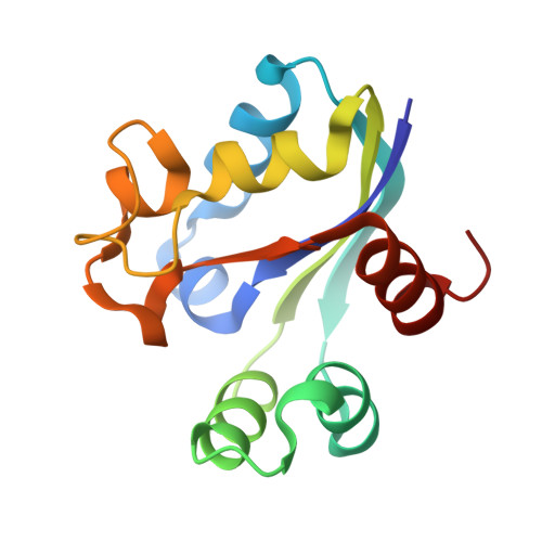
6QA2 - PubMed Abstract:
To be fully active and participate in the metabolism of phosphorylated nucleotides, most nucleoside diphosphate kinases (NDPKs) have to assemble into stable hexamers. Here we studied the role played by six intersubunit salt bridges R80-D93 in the stability of NDPK from the pathogen Mycobacterium tuberculosis ( Mt). Mutating R80 into Ala or Asn abolished the salt bridges. Unexpectedly, compensatory stabilizing mechanisms appeared for R80A and R80N mutants and we studied them by biochemical and structural methods. The R80A mutant crystallized into space group I222 that is unusual for NDPK, and its hexameric structure revealed the occurrence at the trimer interface of a stabilizing hydrophobic patch around the mutation. Functionally relevant, a trimer of the R80A hexamer showed a remodeling of the binding site. In this conformation, the cleft of the active site is more open, and then active His117 is more accessible to substrates. H/D exchange mass spectrometry analysis of the wild type and the R80A and R80N mutants showed that the remodeled region of the protein is highly solvent accessible, indicating that equilibrium between open and closed conformations is possible. We propose that such equilibrium occurs in vivo and explains how bulky substrates access the catalytic His117.
- Université de Bordeaux , CNRS, Institut de Biochimie et Génétique Cellulaires, UMR5095 , 146 rue Léo Saignat , 33077 Bordeaux , France.
Organizational Affiliation: