6Q61 | pdb_00006q61

Pore-modulating toxins exploit inherent slow inactivation to block K+ channels


Experimental Data Snapshot

  • Method: X-RAY DIFFRACTION
  • Resolution: 1.30 Å
  • R-Value Free: 
    0.167 (Depositor), 0.171 (DCC) 
  • R-Value Work: 
    0.139 (Depositor), 0.141 (DCC) 
  • R-Value Observed: 
    0.140 (Depositor) 

Starting Model: experimental
View more details

wwPDB Validation 3D Report Full Report

Validation slider image for 6Q61

Ligand Structure Quality Assessment 


This is version 1.4 of the entry. See complete history

Literature

Pore-modulating toxins exploit inherent slow inactivation to block K+channels.

Karbat, I.Altman-Gueta, H.Fine, S.Szanto, T.Hamer-Rogotner, S.Dym, O.Frolow, F.Gordon, D.Panyi, G.Gurevitz, M.Reuveny, E.

(2019) Proc Natl Acad Sci U S A 116: 18700-18709

  • DOI: https://doi.org/10.1073/pnas.1908903116
  • Primary Citation Related Structures: 
    6Q61, 6Q6C

  • PubMed Abstract: 

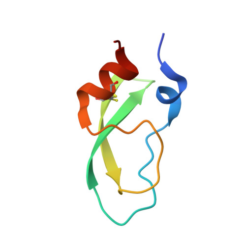
    Voltage-dependent potassium channels (K v s) gate in response to changes in electrical membrane potential by coupling a voltage-sensing module with a K + -selective pore. Animal toxins targeting K v s are classified as pore blockers, which physically plug the ion conduction pathway, or as gating modifiers, which disrupt voltage sensor movements. A third group of toxins blocks K + conduction by an unknown mechanism via binding to the channel turrets. Here, we show that Conkunitzin-S1 (Cs1), a peptide toxin isolated from cone snail venom, binds at the turrets of K v 1.2 and targets a network of hydrogen bonds that govern water access to the peripheral cavities that surround the central pore. The resulting ectopic water flow triggers an asymmetric collapse of the pore by a process resembling that of inherent slow inactivation. Pore modulation by animal toxins exposes the peripheral cavity of K + channels as a novel pharmacological target and provides a rational framework for drug design.


  • Organizational Affiliation
    • Department of Biomolecular Sciences, Weizmann Institute of Science, 76100 Rehovot, Israel.

Macromolecule Content 

  • Total Structure Weight: 7.05 kDa 
  • Atom Count: 568 
  • Modeled Residue Count: 59 
  • Deposited Residue Count: 61 
  • Unique protein chains: 1

Macromolecules

Find similar proteins by:|  3D Structure
Entity ID: 1
MoleculeChains  Sequence LengthOrganismDetailsImage
Kunitz-type conkunitzin-S161Conus striatusMutation(s): 0 
UniProt
Find proteins for P0C1X2 (Conus striatus)
Explore P0C1X2 
Go to UniProtKB:  P0C1X2
Entity Groups
Sequence Clusters30% Identity50% Identity70% Identity90% Identity95% Identity100% Identity
UniProt GroupP0C1X2
Sequence Annotations
Expand
Reference Sequence

Small Molecules

Ligands 1 Unique
IDChains Name / Formula / InChI Key2D Diagram3D Interactions
SO4
(Subject of Investigation/LOI)

Query on SO4


Download:Ideal Coordinates CCD File
B [auth A]SULFATE ION
O4 S
QAOWNCQODCNURD-UHFFFAOYSA-L

Experimental Data & Validation

Experimental Data

  • Method: X-RAY DIFFRACTION
  • Resolution: 1.30 Å
  • R-Value Free:  0.167 (Depositor), 0.171 (DCC) 
  • R-Value Work:  0.139 (Depositor), 0.141 (DCC) 
  • R-Value Observed: 0.140 (Depositor) 
Space Group: P 63
Unit Cell:
Length ( Å )Angle ( ˚ )
a = 51.031α = 90
b = 51.031β = 90
c = 42.569γ = 120
Software Package:
Software NamePurpose
REFMACrefinement
HKL-2000data reduction
HKL-2000data scaling
MOLREPphasing

Structure Validation

View Full Validation Report


Ligand Structure Quality Assessment 


Entry History 

& Funding Information

Deposition Data


Funding OrganizationLocationGrant Number
Israel Science FoundationIsrael1248/2015

Revision History  (Full details and data files)

  • Version 1.0: 2019-08-21
    Type: Initial release
  • Version 1.1: 2019-09-04
    Changes: Data collection, Database references
  • Version 1.2: 2019-09-18
    Changes: Data collection, Database references
  • Version 1.3: 2024-01-24
    Changes: Advisory, Data collection, Database references, Refinement description
  • Version 1.4: 2024-10-23
    Changes: Structure summary