


Tetrahydrofolate Riboswitches Provide Distinct Genetic Outputs to Synthetic and Natural Signals.
Vincent, H.A., Leigh, J., Robinson, C.J., Dunstan, M.S., Ferrer Rios, M.G., Micklefield, J.To be published.
Experimental Data Snapshot
Entity ID: 1 | ||||
| Molecule | Chains | Length | Organism | Image |
|---|---|---|---|---|
| tetrahydrofolate riboswitch aptamer | 89 | Streptococcus mutans UA159 | 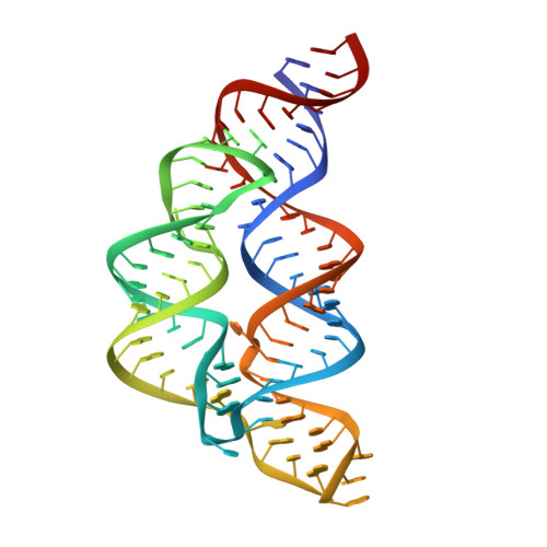 | |
Sequence AnnotationsExpand | ||||
Reference Sequence | ||||
| Ligands 2 Unique | |||||
|---|---|---|---|---|---|
| ID | Chains | Name / Formula / InChI Key | 2D Diagram | 3D Interactions | |
| T0A Download:Ideal Coordinates CCD File | B [auth A], C [auth A] | 5-deazatetrahydropterin C7 H10 N4 O PRQFDDYAWVNJMM-UHFFFAOYSA-N |  | ||
| MG Download:Ideal Coordinates CCD File | D [auth A] | MAGNESIUM ION Mg JLVVSXFLKOJNIY-UHFFFAOYSA-N |  | ||
| Length ( Å ) | Angle ( ˚ ) |
|---|---|
| a = 26.863 | α = 90 |
| b = 68.697 | β = 90 |
| c = 157.309 | γ = 90 |
| Software Name | Purpose |
|---|---|
| PHENIX | refinement |
| PDB_EXTRACT | data extraction |
| xia2 | data reduction |
| xia2 | data scaling |
| PHASER | phasing |
| Funding Organization | Location | Grant Number |
|---|---|---|
| United Kingdom | -- |


