Structure and redox properties of the diheme electron carrier cytochrome c4from Pseudomonas aeruginosa.
Carpenter, J.M., Zhong, F., Ragusa, M.J., Louro, R.O., Hogan, D.A., Pletneva, E.V.(2019) J Inorg Biochem 203: 110889-110889
- PubMed: 31707335 Search on PubMedSearch on PubMed Central
- DOI: https://doi.org/10.1016/j.jinorgbio.2019.110889
- Primary Citation Related Structures:
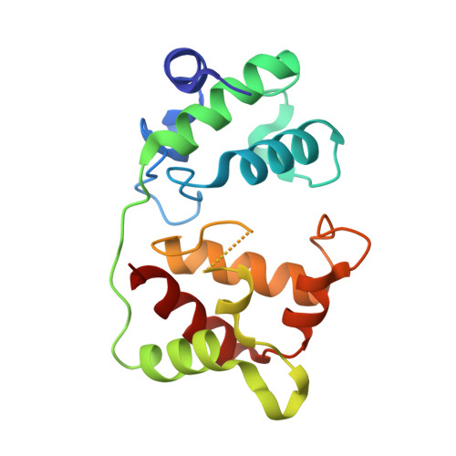
6Q2U - PubMed Abstract:
At low oxygen concentrations, respiration of Pseudomonas aeruginosa (Pa) and other bacteria relies on activity of cytochrome cbb 3 oxidases. A diheme cytochrome c 4 (cyt c 4 ) donates electrons to Pa cbb 3 oxidases to enable oxygen reduction and proton pumping by these enzymes. Given the importance of this redox pathway for bacterial pathogenesis, both cyt c 4 and cbb 3 oxidase are potential targets for new antibacterial strategies. The structural information about these two proteins, however, is scarce, and functional insights for Pa and other bacteria have been primarily drawn from analyses of the analogous system from Pseudomonas stutzeri (Ps). Herein, we describe characterization of structural and redox properties of cyt c 4 from Pa. The crystal structure of Pa cyt c 4 has revealed that this protein is organized in two monoheme domains. The interdomain interface is more hydrophobic in Pa cyt c 4 , and the protein surface does not show the dipolar distribution of charges found in Ps cyt c 4 . The reduction potentials of the two hemes are similar in Pa cyt c 4 but differ by about 100 mV in Ps cyt c 4 . Analyses of structural models of these and other cyt c 4 proteins suggest that multiple factors contribute to the potential difference of the two hemes in these proteins, including solvent accessibility of the heme group, the distribution of surface charges, and the nature of the interdomain interface. The distinct properties of cyt c 4 proteins from closely-related Pa and Ps bacteria emphasize the importance of examining the cbb 3 /cyt c 4 redox pathway in multiple species.
- Department of Chemistry, Dartmouth College, Hanover, NH 03755, United States of America.
Organizational Affiliation: