6PD5 | pdb_00006pd5

Crystal Structure of a H5N1 influenza virus hemagglutinin at pH 6.5


Experimental Data Snapshot

  • Method: X-RAY DIFFRACTION
  • Resolution: 2.39 Å
  • R-Value Free: 
    0.261 (Depositor), 0.259 (DCC) 
  • R-Value Work: 
    0.213 (Depositor), 0.217 (DCC) 
  • R-Value Observed: 
    0.216 (Depositor) 

Starting Model: experimental
View more details

wwPDB Validation 3D Report Full Report

Validation slider image for 6PD5

Ligand Structure Quality Assessment 


This is version 1.5 of the entry. See complete history

Literature

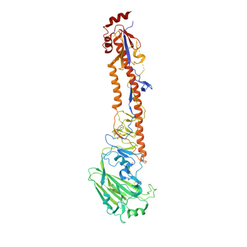
Identification of a pH sensor in Influenza hemagglutinin using X-ray crystallography.

Antanasijevic, A.Durst, M.A.Lavie, A.Caffrey, M.

(2020) J Struct Biol 209: 107412-107412

  • DOI: https://doi.org/10.1016/j.jsb.2019.107412
  • Primary Citation Related Structures: 
    6PCX, 6PD3, 6PD5, 6PD6

  • PubMed Abstract: 

    Hemagglutnin (HA) mediates entry of influenza virus through a series of conformational changes triggered by the low pH of the endosome. The residue or combination of residues acting as pH sensors has not yet been fully elucidated. In this work, we assay pH effects on the structure of H5 HA by soaking HA crystallized at pH 6.5 in a series of buffers with lower pH, mimicking the conditions of the endosome. We find that HA1-H38, which is conserved in Group 1 HA, undergoes a striking change in side chain conformation, which we attribute to its protonation and cation-cation repulsion with conserved HA1-H18. This work suggests that x-ray crystallography can be applied for studying small-scale pH-induced conformational changes providing valuable information on the location of pH sensors in HA. Importantly, the observed change in HA1-H38 conformation is further evidence that the pH-induced conformational changes of HA are the result of a series of protonation events to conserved and non-conserved pH sensors.


  • Organizational Affiliation
    • Department of Biochemistry and Molecular Genetics, University of Illinois at Chicago, 900 S Ashland Ave, 60607 Chicago, USA.

Macromolecule Content 

  • Total Structure Weight: 62.11 kDa 
  • Atom Count: 4,178 
  • Modeled Residue Count: 491 
  • Deposited Residue Count: 527 
  • Unique protein chains: 1

Macromolecules

Find similar proteins by:|  3D Structure
Entity ID: 1
MoleculeChains  Sequence LengthOrganismDetailsImage
Hemagglutinin527Influenza A virus (A/chicken/Vietnam/4/2003(H5N1))Mutation(s): 0 
Gene Names: HA
UniProt
Find proteins for Q1KHJ8 (Influenza A virus)
Explore Q1KHJ8 
Go to UniProtKB:  Q1KHJ8
Entity Groups
Sequence Clusters30% Identity50% Identity70% Identity90% Identity95% Identity100% Identity
UniProt GroupQ1KHJ8
Glycosylation
Glycosylation Sites: 4
Sequence Annotations
Expand
Reference Sequence

Small Molecules

Ligands 3 Unique
IDChains Name / Formula / InChI Key2D Diagram3D Interactions
NAG

Query on NAG


Download:Ideal Coordinates CCD File
P [auth A],
Q [auth A],
R [auth A],
S [auth A]
2-acetamido-2-deoxy-beta-D-glucopyranose
C8 H15 N O6
OVRNDRQMDRJTHS-FMDGEEDCSA-N
SO4

Query on SO4


Download:Ideal Coordinates CCD File
B [auth A]
C [auth A]
D [auth A]
E [auth A]
F [auth A]
B [auth A],
C [auth A],
D [auth A],
E [auth A],
F [auth A],
G [auth A],
H [auth A],
I [auth A],
J [auth A],
K [auth A],
L [auth A]
SULFATE ION
O4 S
QAOWNCQODCNURD-UHFFFAOYSA-L
GOL

Query on GOL


Download:Ideal Coordinates CCD File
M [auth A],
N [auth A],
O [auth A]
GLYCEROL
C3 H8 O3
PEDCQBHIVMGVHV-UHFFFAOYSA-N

Experimental Data & Validation

Experimental Data

  • Method: X-RAY DIFFRACTION
  • Resolution: 2.39 Å
  • R-Value Free:  0.261 (Depositor), 0.259 (DCC) 
  • R-Value Work:  0.213 (Depositor), 0.217 (DCC) 
  • R-Value Observed: 0.216 (Depositor) 
Space Group: H 3 2
Unit Cell:
Length ( Å )Angle ( ˚ )
a = 108.056α = 90
b = 108.056β = 90
c = 419.819γ = 120
Software Package:
Software NamePurpose
REFMACrefinement
XDSdata reduction
XDSdata scaling
MOLREPphasing
Cootmodel building
PDB_EXTRACTdata extraction

Structure Validation

View Full Validation Report


Ligand Structure Quality Assessment 


Entry History 

Deposition Data

Revision History  (Full details and data files)

  • Version 1.0: 2019-11-13
    Type: Initial release
  • Version 1.1: 2019-11-20
    Changes: Database references
  • Version 1.2: 2020-01-22
    Changes: Database references
  • Version 1.3: 2020-07-29
    Type: Remediation
    Reason: Carbohydrate remediation
    Changes: Data collection, Derived calculations, Structure summary
  • Version 1.4: 2023-10-11
    Changes: Data collection, Database references, Refinement description, Structure summary
  • Version 1.5: 2024-11-20
    Changes: Structure summary