6OZH

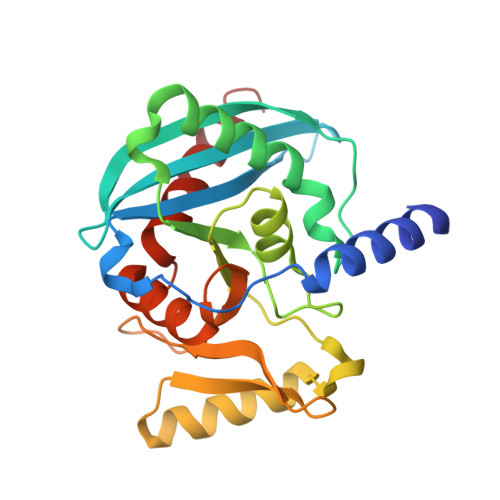
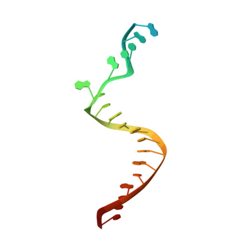
Crystal structure of Ciona intestinalis (Ci) Endonuclease V in complex with a 24mer DNA containing an inosine followed by a ribo-adenosine


Experimental Data Snapshot

  • Method: X-RAY DIFFRACTION
  • Resolution: 3.03 Å
  • R-Value Free: 0.269 
  • R-Value Work: 0.233 
  • R-Value Observed: 0.235 

wwPDB Validation   3D Report Full Report


This is version 1.2 of the entry. See complete history


Literature

Evolution of Inosine-Specific Endonuclease V from Bacterial DNase to Eukaryotic RNase.

Wu, J.Samara, N.L.Kuraoka, I.Yang, W.

(2019) Mol Cell 76: 44

  • DOI: https://doi.org/10.1016/j.molcel.2019.06.046
  • Primary Citation of Related Structures:  
    6OZE, 6OZF, 6OZG, 6OZH, 6OZI, 6OZJ, 6OZK, 6OZL, 6OZM, 6OZN, 6OZO, 6OZP, 6OZQ, 6OZR, 6OZS

  • PubMed Abstract: 

    Endonuclease V (EndoV) cleaves the second phosphodiester bond 3' to a deaminated adenosine (inosine). Although highly conserved, EndoV homologs change substrate preference from DNA in bacteria to RNA in eukaryotes. We have characterized EndoV from six different species and determined crystal structures of human EndoV and three EndoV homologs from bacteria to mouse in complex with inosine-containing DNA/RNA hybrid or double-stranded RNA (dsRNA). Inosine recognition is conserved, but changes in several connecting loops in eukaryotic EndoV confer recognition of 3 ribonucleotides upstream and 7 or 8 bp of dsRNA downstream of the cleavage site, and bacterial EndoV binds only 2 or 3 nt flanking the scissile phosphate. In addition to the two canonical metal ions in the active site, a third Mn 2+ that coordinates the nucleophilic water appears necessary for product formation. Comparison of EndoV with its homologs RNase H1 and Argonaute reveals the principles by which these enzymes recognize RNA versus DNA.


  • Organizational Affiliation

    Laboratory of Molecular Biology, NIDDK, NIH, Bethesda, MD 20892, USA.


Macromolecules

Find similar proteins by:  (by identity cutoff)  |  3D Structure
Entity ID: 1
MoleculeChains Sequence LengthOrganismDetailsImage
endonuclease V isoform X2
A, B, C, D
245Ciona intestinalisMutation(s): 0 
Gene Names: LOC100181026
Entity Groups  
Sequence Clusters30% Identity50% Identity70% Identity90% Identity95% Identity100% Identity
Sequence Annotations
Expand
  • Reference Sequence

Find similar nucleic acids by:  Sequence   |   3D Structure  

Entity ID: 2
MoleculeChains LengthOrganismImage
DNA/RNA (5'-D(P*CP*GP*GP*TP*AP*AP*CP*CP*GP*I)-R(P*A)-D(P*TP*AP*TP*GP*CP*AP*GP*CP*AP*TP*TP*TP*C)-3')
E, F, G, H
24synthetic construct
Sequence Annotations
Expand
  • Reference Sequence
Experimental Data & Validation

Experimental Data

  • Method: X-RAY DIFFRACTION
  • Resolution: 3.03 Å
  • R-Value Free: 0.269 
  • R-Value Work: 0.233 
  • R-Value Observed: 0.235 
  • Space Group: P 1 21 1
Unit Cell:
Length ( Å )Angle ( ˚ )
a = 74.245α = 90
b = 121.975β = 98.75
c = 79.921γ = 90
Software Package:
Software NamePurpose
PHENIXrefinement
HKL-2000data reduction
HKL-2000data scaling
PHENIXphasing

Structure Validation

View Full Validation Report


Entry History 

Deposition Data

Revision History  (Full details and data files)

  • Version 1.0: 2019-09-04
    Type: Initial release
  • Version 1.1: 2019-10-16
    Changes: Data collection, Database references
  • Version 1.2: 2024-03-13
    Changes: Data collection, Database references, Derived calculations