6OYA

Structure of the Rhodopsin-Transducin-Nanobody Complex


Experimental Data Snapshot

  • Method: ELECTRON MICROSCOPY
  • Resolution: 3.30 Å
  • Aggregation State: PARTICLE 
  • Reconstruction Method: SINGLE PARTICLE 

wwPDB Validation   3D Report Full Report


This is version 1.3 of the entry. See complete history


Literature

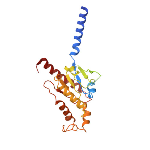
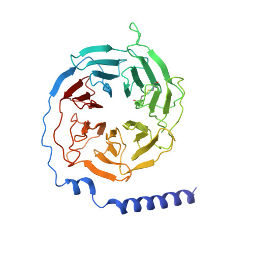
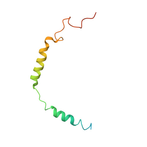
Structures of the Rhodopsin-Transducin Complex: Insights into G-Protein Activation.

Gao, Y.Hu, H.Ramachandran, S.Erickson, J.W.Cerione, R.A.Skiniotis, G.

(2019) Mol Cell 75: 781

  • DOI: https://doi.org/10.1016/j.molcel.2019.06.007
  • Primary Citation of Related Structures:  
    6OY9, 6OYA

  • PubMed Abstract: 
  • Rhodopsin (Rho), a prototypical G-protein-coupled receptor (GPCR) in vertebrate vision, activates the G-protein transducin (G T ) by catalyzing GDP-GTP exchange on its α subunit (Gα T ). To elucidate the determinants of G T coupling and activation, we obtained cryo-EM structures of a fully functional, light-activated Rho-G T complex in the presence and absence of a G-protein-stabilizing nanobody ...

    Rhodopsin (Rho), a prototypical G-protein-coupled receptor (GPCR) in vertebrate vision, activates the G-protein transducin (G T ) by catalyzing GDP-GTP exchange on its α subunit (Gα T ). To elucidate the determinants of G T coupling and activation, we obtained cryo-EM structures of a fully functional, light-activated Rho-G T complex in the presence and absence of a G-protein-stabilizing nanobody. The structures illustrate how G T overcomes its low basal activity by engaging activated Rho in a conformation distinct from other GPCR-G-protein complexes. Moreover, the nanobody-free structures reveal native conformations of G-protein components and capture three distinct conformers showing the Gα T helical domain (αHD) contacting the Gβγ subunits. These findings uncover the molecular underpinnings of G-protein activation by visual rhodopsin and shed new light on the role played by Gβγ during receptor-catalyzed nucleotide exchange.


    Organizational Affiliation

    Department of Structural Biology, Stanford University School of Medicine, Stanford, CA 94305, USA; Department of Molecular and Cellular Physiology, Stanford University School of Medicine, Stanford, CA 94305, USA. Electronic address: yiorgo@stanford.edu.


Macromolecules
Find similar proteins by:  (by identity cutoff)  |  3D Structure
Entity ID: 1
MoleculeChainsSequence LengthOrganismDetailsImage
Gt-alpha/Gi1-alpha chimeraA359Bos taurusMutation(s): 0 
Gene Names: GNAT1
Membrane Entity: Yes 
UniProt
Find proteins for P04695 (Bos taurus)
Explore P04695 
Go to UniProtKB:  P04695
Entity Groups  
Sequence Clusters30% Identity50% Identity70% Identity90% Identity95% Identity100% Identity
UniProt GroupP04695
Protein Feature View
Expand
  • Reference Sequence
Find similar proteins by:  (by identity cutoff)  |  3D Structure
Entity ID: 2
MoleculeChainsSequence LengthOrganismDetailsImage
Guanine nucleotide-binding protein G(I)/G(S)/G(T) subunit beta-1B340Bos taurusMutation(s): 0 
Gene Names: GNB1
UniProt
Find proteins for P62871 (Bos taurus)
Explore P62871 
Go to UniProtKB:  P62871
Entity Groups  
Sequence Clusters30% Identity50% Identity70% Identity90% Identity95% Identity100% Identity
UniProt GroupP62871
Protein Feature View
Expand
  • Reference Sequence
Find similar proteins by:  (by identity cutoff)  |  3D Structure
Entity ID: 3
MoleculeChainsSequence LengthOrganismDetailsImage
Guanine nucleotide-binding protein G(T) subunit gamma-T1C [auth G]81Bos taurusMutation(s): 0 
Gene Names: GNGT1
Membrane Entity: Yes 
UniProt
Find proteins for P02698 (Bos taurus)
Explore P02698 
Go to UniProtKB:  P02698
Entity Groups  
Sequence Clusters30% Identity50% Identity70% Identity90% Identity95% Identity100% Identity
UniProt GroupP02698
Protein Feature View
Expand
  • Reference Sequence
Find similar proteins by:  (by identity cutoff)  |  3D Structure
Entity ID: 4
MoleculeChainsSequence LengthOrganismDetailsImage
Camelid antibody VHH fragmentD [auth N]138Lama glamaMutation(s): 0 
Entity Groups  
Sequence Clusters30% Identity50% Identity70% Identity90% Identity95% Identity100% Identity
Protein Feature View
Expand
  • Reference Sequence
Find similar proteins by:  (by identity cutoff)  |  3D Structure
Entity ID: 5
MoleculeChainsSequence LengthOrganismDetailsImage
RhodopsinE [auth R]348Bos taurusMutation(s): 0 
Gene Names: RHO
Membrane Entity: Yes 
UniProt
Find proteins for P02699 (Bos taurus)
Explore P02699 
Go to UniProtKB:  P02699
Entity Groups  
Sequence Clusters30% Identity50% Identity70% Identity90% Identity95% Identity100% Identity
UniProt GroupP02699
Protein Feature View
Expand
  • Reference Sequence
Small Molecules
Ligands 1 Unique
IDChainsName / Formula / InChI Key2D Diagram3D Interactions
RET (Subject of Investigation/LOI)
Query on RET

Download Ideal Coordinates CCD File 
F [auth R]RETINAL
C20 H28 O
NCYCYZXNIZJOKI-OVSJKPMPSA-N
 Ligand Interaction
Experimental Data & Validation

Experimental Data

  • Method: ELECTRON MICROSCOPY
  • Resolution: 3.30 Å
  • Aggregation State: PARTICLE 
  • Reconstruction Method: SINGLE PARTICLE 
EM Software:
TaskSoftware PackageVersion
RECONSTRUCTIONcisTEM

Structure Validation

View Full Validation Report


Entry History & Funding Information

Deposition Data


Funding OrganizationLocationGrant Number
National Institutes of Health/National Cancer Institute (NIH/NCI)United StatesR01 CA201402
National Institutes of Health/National Institute of General Medical Sciences (NIH/NIGMS)United StatesR35 GM122575
National Institutes of Health/National Institute of Neurological Disorders and Stroke (NIH/NINDS)United StatesR01 NS092695

Revision History  (Full details and data files)

  • Version 1.0: 2019-07-24
    Type: Initial release
  • Version 1.1: 2019-09-04
    Changes: Data collection, Database references
  • Version 1.2: 2019-11-27
    Changes: Data collection
  • Version 1.3: 2019-12-04
    Changes: Author supporting evidence