6OX3

SETD3 in Complex with an Actin Peptide with His73 Replaced with Lysine


Experimental Data Snapshot

  • Method: X-RAY DIFFRACTION
  • Resolution: 1.78 Å
  • R-Value Free: 0.246 
  • R-Value Work: 0.208 
  • R-Value Observed: 0.208 

wwPDB Validation   3D Report Full Report


Ligand Structure Quality Assessment 


This is version 1.1 of the entry. See complete history


Literature

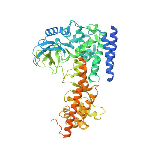
Structural basis for the target specificity of actin histidine methyltransferase SETD3.

Dai, S.Horton, J.R.Woodcock, C.B.Wilkinson, A.W.Zhang, X.Gozani, O.Cheng, X.

(2019) Nat Commun 10: 3541-3541

  • DOI: 10.1038/s41467-019-11554-6
  • Primary Citation of Related Structures:  
    6OX0, 6OX1, 6OX2, 6OX3, 6OX4, 6OX5

  • PubMed Abstract: 
  • SETD3 is an actin histidine-N 3 methyltransferase, whereas other characterized SET-domain enzymes are protein lysine methyltransferases. We report that in a pre-reactive complex SETD3 binds the N 3 -protonated form (N 3 -H) of actin His73, and in a post-reactive product complex, SETD3 generates the methylated histidine in an N 1 -protonated (N 1 -H) and N 3 -methylated form ...

    SETD3 is an actin histidine-N 3 methyltransferase, whereas other characterized SET-domain enzymes are protein lysine methyltransferases. We report that in a pre-reactive complex SETD3 binds the N 3 -protonated form (N 3 -H) of actin His73, and in a post-reactive product complex, SETD3 generates the methylated histidine in an N 1 -protonated (N 1 -H) and N 3 -methylated form. During the reaction, the imidazole ring of His73 rotates ~105°, which shifts the proton from N 3 to N 1 , thus ensuring that the target atom N 3 is deprotonated prior to the methyl transfer. Under the conditions optimized for lysine deprotonation, SETD3 has weak lysine methylation activity on an actin peptide in which the target His73 is substituted by a lysine. The structure of SETD3 with Lys73-containing peptide reveals a bent conformation of Lys73, with its side chain aliphatic carbons tracing along the edge of imidazole ring and the terminal ε-amino group occupying a position nearly identical to the N 3 atom of unmethylated histidine.


    Related Citations: 
    • SETD3 is an actin histidine methyltransferase that prevents primary dystocia.
      Wilkinson, A.W., Diep, J., Dai, S., Liu, S., Ooi, Y.S., Song, D., Li, T.M., Horton, J.R., Zhang, X., Liu, C., Trivedi, D.V., Ruppel, K.M., Vilches-Moure, J.G., Casey, K.M., Mak, J., Cowan, T., Elias, J.E., Nagamine, C.M., Spudich, J.A., Cheng, X., Carette, J.E., Gozani, O.
      (2019) Nature 565: 372

    Organizational Affiliation

    Department of Epigenetics and Molecular Carcinogenesis, The University of Texas MD Anderson Cancer Center, Houston, TX, 77030, USA. xcheng5@mdanderson.org.


Macromolecules

Find similar proteins by:  Sequence   |   3D Structure  

Entity ID: 1
MoleculeChainsSequence LengthOrganismDetailsImage
Actin PeptideA [auth Y],
C [auth Z]
19Homo sapiensMutation(s): 1 
Gene Names: ACTB
UniProt & NIH Common Fund Data Resources
Find proteins for P60709 (Homo sapiens)
Explore P60709 
Go to UniProtKB:  P60709
PHAROS:  P60709
Entity Groups  
Sequence Clusters30% Identity50% Identity70% Identity90% Identity95% Identity100% Identity
UniProt GroupP60709
Protein Feature View
Expand
  • Reference Sequence
Find similar proteins by:  (by identity cutoff)  |  3D Structure
Entity ID: 2
MoleculeChainsSequence LengthOrganismDetailsImage
Histone-lysine N-methyltransferase setd3B [auth A],
D [auth B]
599Homo sapiensMutation(s): 0 
Gene Names: SETD3C14orf154
EC: 2.1.1.85
UniProt & NIH Common Fund Data Resources
Find proteins for Q86TU7 (Homo sapiens)
Explore Q86TU7 
Go to UniProtKB:  Q86TU7
PHAROS:  Q86TU7
Entity Groups  
Sequence Clusters30% Identity50% Identity70% Identity90% Identity95% Identity100% Identity
UniProt GroupQ86TU7
Protein Feature View
Expand
  • Reference Sequence
Small Molecules
Ligands 5 Unique
IDChainsName / Formula / InChI Key2D Diagram3D Interactions
SAH
Query on SAH

Download Ideal Coordinates CCD File 
F [auth A],
HA [auth B]
S-ADENOSYL-L-HOMOCYSTEINE
C14 H20 N6 O5 S
ZJUKTBDSGOFHSH-WFMPWKQPSA-N
 Ligand Interaction
PGE
Query on PGE

Download Ideal Coordinates CCD File 
UA [auth B]TRIETHYLENE GLYCOL
C6 H14 O4
ZIBGPFATKBEMQZ-UHFFFAOYSA-N
 Ligand Interaction
GOL
Query on GOL

Download Ideal Coordinates CCD File 
E [auth Y],
G [auth A],
H [auth A],
I [auth A],
J [auth A]
GLYCEROL
C3 H8 O3
PEDCQBHIVMGVHV-UHFFFAOYSA-N
 Ligand Interaction
EDO
Query on EDO

Download Ideal Coordinates CCD File 
AA [auth A],
BA [auth A],
CA [auth A],
DA [auth A],
EA [auth A],
AA [auth A],
BA [auth A],
CA [auth A],
DA [auth A],
EA [auth A],
FA [auth A],
IA [auth B],
JA [auth B],
K [auth A],
KA [auth B],
L [auth A],
LA [auth B],
M [auth A],
MA [auth B],
N [auth A],
NA [auth B],
O [auth A],
OA [auth B],
P [auth A],
PA [auth B],
Q [auth A],
QA [auth B],
R [auth A],
RA [auth B],
S [auth A],
SA [auth B],
T [auth A],
TA [auth B],
U [auth A],
V [auth A],
W [auth A],
X [auth A],
Y [auth A],
Z [auth A]
1,2-ETHANEDIOL
C2 H6 O2
LYCAIKOWRPUZTN-UHFFFAOYSA-N
 Ligand Interaction
ACT
Query on ACT

Download Ideal Coordinates CCD File 
GA [auth A]ACETATE ION
C2 H3 O2
QTBSBXVTEAMEQO-UHFFFAOYSA-M
 Ligand Interaction
Experimental Data & Validation

Experimental Data

  • Method: X-RAY DIFFRACTION
  • Resolution: 1.78 Å
  • R-Value Free: 0.246 
  • R-Value Work: 0.208 
  • R-Value Observed: 0.208 
  • Space Group: P 1 21 1
Unit Cell:
Length ( Å )Angle ( ˚ )
a = 60.233α = 90
b = 175.748β = 92.39
c = 66.471γ = 90
Software Package:
Software NamePurpose
PHENIXrefinement
HKL-2000data reduction
HKL-2000data scaling
PHENIXphasing

Structure Validation

View Full Validation Report


Ligand Structure Quality Assessment 


Entry History & Funding Information

Deposition Data


Funding OrganizationLocationGrant Number
National Institutes of Health/National Institute of General Medical Sciences (NIH/NIGMS)United StatesGM049245-23

Revision History  (Full details and data files)

  • Version 1.0: 2019-08-21
    Type: Initial release
  • Version 1.1: 2020-01-01
    Changes: Author supporting evidence