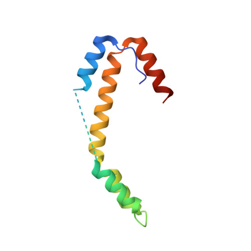
6OWZ

Spy H96L:Im7 L19pI-Phe complex; multiple anomalous datasets contained herein for element identification


Experimental Data Snapshot

  • Method: X-RAY DIFFRACTION
  • Resolution: 2.05 Å
  • R-Value Free: 0.255 
  • R-Value Work: 0.215 
  • R-Value Observed: 0.217 

Starting Model: experimental
View more details

wwPDB Validation   3D Report Full Report


This is version 2.3 of the entry. See complete history


Literature

Identifying dynamic, partially occupied residues using anomalous scattering.

Rocchio, S.Duman, R.El Omari, K.Mykhaylyk, V.Orr, C.Yan, Z.Salmon, L.Wagner, A.Bardwell, J.C.A.Horowitz, S.

(2019) Acta Crystallogr D Struct Biol 75: 1084-1095

  • DOI: https://doi.org/10.1107/S2059798319014475
  • Primary Citation of Related Structures:  
    6OWX, 6OWY, 6OWZ

  • PubMed Abstract: 

    Although often presented as taking single `snapshots' of the conformation of a protein, X-ray crystallography provides an averaged structure over time and space within the crystal. The important but difficult task of characterizing structural ensembles in crystals is typically limited to small conformational changes, such as multiple side-chain conformations. A crystallographic method was recently introduced that utilizes residual electron and anomalous density (READ) to characterize structural ensembles encompassing large-scale structural changes. Key to this method is an ability to accurately measure anomalous signals and distinguish them from noise or other anomalous scatterers. This report presents an optimized data-collection and analysis strategy for partially occupied iodine anomalous signals. Using the long-wavelength-optimized beamline I23 at Diamond Light Source, the ability to accurately distinguish the positions of anomalous scatterers with occupancies as low as ∼12% is demonstrated. The number and positions of these anomalous scatterers are consistent with previous biophysical, kinetic and structural data that suggest that the protein Im7 binds to the chaperone Spy in multiple partially occupied conformations. Finally, READ selections demonstrate that re-measured data using the new protocols are consistent with the previously characterized structural ensemble of the chaperone Spy with its client Im7. This study shows that a long-wavelength beamline results in easily validated anomalous signals that are strong enough to be used to detect and characterize highly disordered sections of crystal structures.


  • Organizational Affiliation

    Department of Molecular, Cellular and Developmental Biology, and the Howard Hughes Medical Institute, University of Michigan, Ann Arbor, MI 48109, USA.


Macromolecules
Find similar proteins by:  (by identity cutoff)  |  3D Structure
Entity ID: 1
MoleculeChains Sequence LengthOrganismDetailsImage
Periplasmic chaperone Spy
A, B
97Escherichia coliMutation(s): 1 
UniProt
Find proteins for P77754 (Escherichia coli (strain K12))
Explore P77754 
Go to UniProtKB:  P77754
Entity Groups  
Sequence Clusters30% Identity50% Identity70% Identity90% Identity95% Identity100% Identity
UniProt GroupP77754
Sequence Annotations
Expand
  • Reference Sequence
Small Molecules
Ligands 4 Unique
IDChains Name / Formula / InChI Key2D Diagram3D Interactions
IOD (Subject of Investigation/LOI)
Query on IOD

Download Ideal Coordinates CCD File 
P [auth A],
Q [auth A],
U [auth B],
V [auth B]
IODIDE ION
I
XMBWDFGMSWQBCA-UHFFFAOYSA-M
IMD
Query on IMD

Download Ideal Coordinates CCD File 
N [auth A],
O [auth A],
T [auth B]
IMIDAZOLE
C3 H5 N2
RAXXELZNTBOGNW-UHFFFAOYSA-O
ZN
Query on ZN

Download Ideal Coordinates CCD File 
C [auth A]
D [auth A]
E [auth A]
F [auth A]
G [auth A]
C [auth A],
D [auth A],
E [auth A],
F [auth A],
G [auth A],
H [auth A],
I [auth A],
J [auth A],
K [auth A],
R [auth B],
S [auth B]
ZINC ION
Zn
PTFCDOFLOPIGGS-UHFFFAOYSA-N
CL
Query on CL

Download Ideal Coordinates CCD File 
L [auth A],
M [auth A]
CHLORIDE ION
Cl
VEXZGXHMUGYJMC-UHFFFAOYSA-M
Experimental Data & Validation

Experimental Data

Unit Cell:
Length ( Å )Angle ( ˚ )
a = 43.02α = 90
b = 43.02β = 90
c = 257.15γ = 90
Software Package:
Software NamePurpose
PHENIXrefinement
XDSdata reduction
Aimlessdata scaling
PHENIXphasing

Structure Validation

View Full Validation Report


Entry History & Funding Information

Deposition Data


Funding OrganizationLocationGrant Number
National Institutes of Health/National Institute of General Medical Sciences (NIH/NIGMS)United StatesR00 GM120388
Howard Hughes Medical Institute (HHMI)United States--

Revision History  (Full details and data files)

  • Version 1.0: 2019-05-29
    Type: Initial release
  • Version 1.1: 2019-06-05
    Changes: Data collection, Database references, Structure summary
  • Version 2.0: 2019-10-09
    Type: Coordinate replacement
    Reason: Atoms with unrealistic or zero occupancies
    Changes: Advisory, Atomic model, Author supporting evidence, Data collection, Derived calculations, Other, Refinement description, Structure summary
  • Version 2.1: 2019-11-20
    Changes: Author supporting evidence
  • Version 2.2: 2019-12-11
    Changes: Database references
  • Version 2.3: 2023-10-11
    Changes: Data collection, Database references, Derived calculations, Refinement description