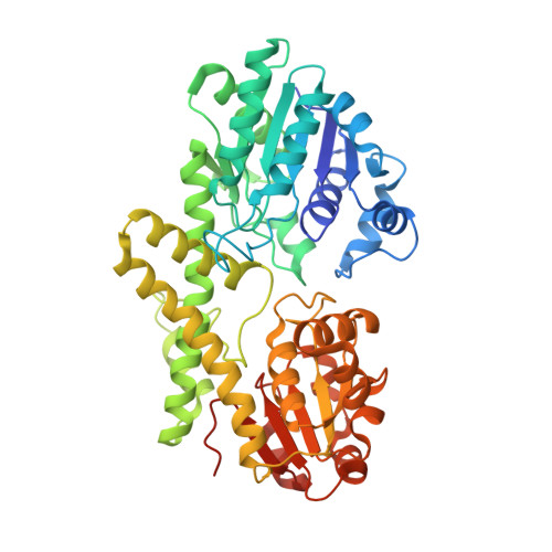
6OM8

Caenorhabditis Elegans UDP-Glucose Dehydrogenase in complex with UDP-Xylose


Experimental Data Snapshot

  • Method: X-RAY DIFFRACTION
  • Resolution: 2.45 Å
  • R-Value Free: 0.220 
  • R-Value Work: 0.194 
  • R-Value Observed: 0.194 

wwPDB Validation   3D Report Full Report


Ligand Structure Quality Assessment 


This is version 1.3 of the entry. See complete history


Literature

Conservation of Atypical Allostery inC. elegansUDP-Glucose Dehydrogenase.

Beattie, N.R.Keul, N.D.Hicks Sirmans, T.N.McDonald, W.E.Talmadge, T.M.Taujale, R.Kannan, N.Wood, Z.A.

(2019) ACS Omega 4: 16318-16329

  • DOI: https://doi.org/10.1021/acsomega.9b01565
  • Primary Citation of Related Structures:  
    6OM8

  • PubMed Abstract: 

    Human UDP-glucose dehydrogenase (hUGDH) oxidizes uridine diphosphate (UDP)-glucose to UDP-glucuronic acid, an essential substrate in the phase II metabolism of drugs. The activity of hUGDH is controlled by an atypical allosteric mechanism in which the feedback inhibitor UDP-xylose competes with the substrate for the active site and triggers a buried allosteric switch to produce an inactive complex (E Ω ). Previous comparisons with a nonallosteric UGDH identified six large-to-small substitutions that produce packing defects in the protein core and provide the conformational flexibility necessary for the allosteric transition. Here, we test the hypothesis that these large-to-small substitutions form a motif that can be used to identify allosteric UGDHs. Caenorhabditis elegans UGDH (cUGDH) conserves this motif with the exception of an Ala-to-Pro substitution in position 109. The crystal structures of unliganded and UDP-xylose bound cUGDH show that the A109P substitution is accommodated by an Asn-to-Ser substitution at position 290. Steady-state analysis and sedimentation velocity studies show that the allosteric transition is conserved in cUGDH. The enzyme also exhibits hysteresis in progress curves and negative cooperativity with respect to NAD + binding. Both of these phenomena are conserved in the human enzyme, which is strong evidence that these represent fundamental features of atypical allostery in UGDH. A phylogenetic analysis of UGDH shows that the atypical allostery motif is ancient and identifies a potential transition point in the evolution of the UGDH family.


  • Organizational Affiliation

    Department of Biochemistry & Molecular Biology and Institute of Bioinformatics, University of Georgia, Athens, Georgia 30602, United States.


Macromolecules
Find similar proteins by:  (by identity cutoff)  |  3D Structure
Entity ID: 1
MoleculeChains Sequence LengthOrganismDetailsImage
UDP-glucose 6-dehydrogenase
A, B, C, D, E
A, B, C, D, E, F, G, H, I, J, K, L
481Caenorhabditis elegansMutation(s): 0 
Gene Names: sqv-4F29F11.1
EC: 1.1.1.22
UniProt
Find proteins for Q19905 (Caenorhabditis elegans)
Explore Q19905 
Go to UniProtKB:  Q19905
Entity Groups  
Sequence Clusters30% Identity50% Identity70% Identity90% Identity95% Identity100% Identity
UniProt GroupQ19905
Sequence Annotations
Expand
  • Reference Sequence
Small Molecules
Ligands 1 Unique
IDChains Name / Formula / InChI Key2D Diagram3D Interactions
UDX
Query on UDX

Download Ideal Coordinates CCD File 
AA [auth H]
BA [auth H]
CA [auth I]
DA [auth I]
EA [auth J]
AA [auth H],
BA [auth H],
CA [auth I],
DA [auth I],
EA [auth J],
FA [auth J],
GA [auth K],
HA [auth K],
IA [auth L],
JA [auth L],
M [auth A],
N [auth A],
O [auth B],
P [auth B],
Q [auth C],
R [auth C],
S [auth D],
T [auth D],
U [auth E],
V [auth E],
W [auth F],
X [auth F],
Y [auth G],
Z [auth G]
URIDINE-5'-DIPHOSPHATE-XYLOPYRANOSE
C14 H22 N2 O16 P2
DQQDLYVHOTZLOR-OCIMBMBZSA-N
Experimental Data & Validation

Experimental Data

  • Method: X-RAY DIFFRACTION
  • Resolution: 2.45 Å
  • R-Value Free: 0.220 
  • R-Value Work: 0.194 
  • R-Value Observed: 0.194 
  • Space Group: P 2 21 21
Unit Cell:
Length ( Å )Angle ( ˚ )
a = 157.71α = 90
b = 168.17β = 90
c = 279.56γ = 90
Software Package:
Software NamePurpose
PHENIXrefinement
XDSdata scaling
Cootmodel building
PHENIXphasing
XDSdata reduction

Structure Validation

View Full Validation Report


Ligand Structure Quality Assessment 


Entry History & Funding Information

Deposition Data


Funding OrganizationLocationGrant Number
National Institutes of Health/National Institute of General Medical Sciences (NIH/NIGMS)United StatesR01GM11429

Revision History  (Full details and data files)

  • Version 1.0: 2019-05-22
    Type: Initial release
  • Version 1.1: 2019-10-30
    Changes: Data collection, Database references
  • Version 1.2: 2020-01-01
    Changes: Author supporting evidence
  • Version 1.3: 2023-10-11
    Changes: Data collection, Database references, Refinement description