6OHU

Structure of EBP and tamoxifen


Experimental Data Snapshot

  • Method: X-RAY DIFFRACTION
  • Resolution: 3.53 Å
  • R-Value Free: 0.326 
  • R-Value Work: 0.273 
  • R-Value Observed: 0.278 

wwPDB Validation   3D Report Full Report


Ligand Structure Quality Assessment 


This is version 1.1 of the entry. See complete history


Literature

Structural basis for human sterol isomerase in cholesterol biosynthesis and multidrug recognition.

Long, T.Hassan, A.Thompson, B.M.McDonald, J.G.Wang, J.Li, X.

(2019) Nat Commun 10: 2452-2452

  • DOI: https://doi.org/10.1038/s41467-019-10279-w
  • Primary Citation of Related Structures:  
    6OHT, 6OHU

  • PubMed Abstract: 

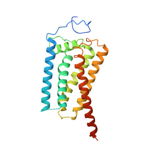
    3-β-hydroxysteroid-Δ 8 , Δ 7 -isomerase, known as Emopamil-Binding Protein (EBP), is an endoplasmic reticulum membrane protein involved in cholesterol biosynthesis, autophagy, oligodendrocyte formation. The mutation on EBP can cause Conradi-Hunermann syndrome, an inborn error. Interestingly, EBP binds an abundance of structurally diverse pharmacologically active compounds, causing drug resistance. Here, we report two crystal structures of human EBP, one in complex with the anti-breast cancer drug tamoxifen and the other in complex with the cholesterol biosynthesis inhibitor U18666A. EBP adopts an unreported fold involving five transmembrane-helices (TMs) that creates a membrane cavity presenting a pharmacological binding site that accommodates multiple different ligands. The compounds exploit their positively-charged amine group to mimic the carbocationic sterol intermediate. Mutagenesis studies on specific residues abolish the isomerase activity and decrease the multidrug binding capacity. This work reveals the catalytic mechanism of EBP-mediated isomerization in cholesterol biosynthesis and how this protein may act as a multi-drug binder.


  • Organizational Affiliation

    Department of Molecular Genetics, University of Texas Southwestern Medical Center, Dallas, TX, 75390, USA.


Macromolecules
Find similar proteins by:  (by identity cutoff)  |  3D Structure
Entity ID: 1
MoleculeChains Sequence LengthOrganismDetailsImage
3-beta-hydroxysteroid-Delta(8),Delta(7)-isomerase
A, B, C
238Homo sapiensMutation(s): 0 
Gene Names: EBP
EC: 5.3.3.5
Membrane Entity: Yes 
UniProt & NIH Common Fund Data Resources
Find proteins for Q15125 (Homo sapiens)
Explore Q15125 
Go to UniProtKB:  Q15125
PHAROS:  Q15125
GTEx:  ENSG00000147155 
Entity Groups  
Sequence Clusters30% Identity50% Identity70% Identity90% Identity95% Identity100% Identity
UniProt GroupQ15125
Sequence Annotations
Expand
  • Reference Sequence
Small Molecules
Binding Affinity Annotations 
IDSourceBinding Affinity
CTX BindingDB:  6OHU Ki: 5 (nM) from 1 assay(s)
IC50: 12 (nM) from 1 assay(s)
Experimental Data & Validation

Experimental Data

  • Method: X-RAY DIFFRACTION
  • Resolution: 3.53 Å
  • R-Value Free: 0.326 
  • R-Value Work: 0.273 
  • R-Value Observed: 0.278 
  • Space Group: P 21 21 2
Unit Cell:
Length ( Å )Angle ( ˚ )
a = 192.837α = 90
b = 66.829β = 90
c = 98.678γ = 90
Software Package:
Software NamePurpose
PHENIXrefinement
HKL-3000data reduction
HKL-3000data scaling
PHENIXphasing

Structure Validation

View Full Validation Report


Ligand Structure Quality Assessment 


Entry History 

Deposition Data

Revision History  (Full details and data files)

  • Version 1.0: 2019-06-19
    Type: Initial release
  • Version 1.1: 2024-03-13
    Changes: Data collection, Database references