Unified Model for Photophysical and Electro-Optical Properties of Green Fluorescent Proteins.
Lin, C.Y., Romei, M.G., Oltrogge, L.M., Mathews, I.I., Boxer, S.G.(2019) J Am Chem Soc 141: 15250-15265
- PubMed: 31450887 Search on PubMedSearch on PubMed Central
- DOI: https://doi.org/10.1021/jacs.9b07152
- Primary Citation Related Structures:
6OFK, 6OFL, 6OFM, 6OFN, 6OFO - PubMed Abstract:
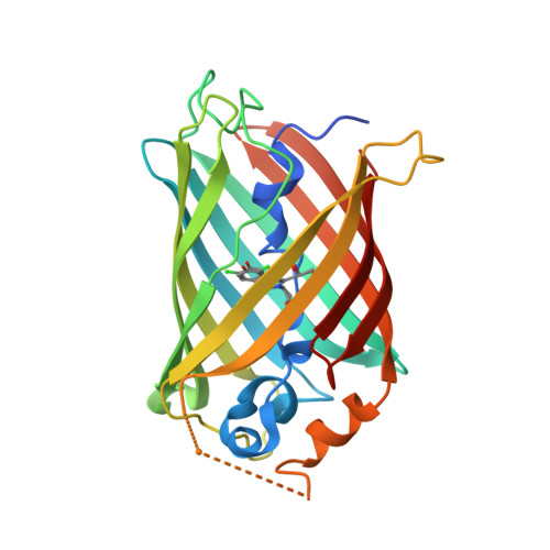
Green fluorescent proteins (GFPs) have become indispensable imaging and optogenetic tools. Their absorption and emission properties can be optimized for specific applications. Currently, no unified framework exists to comprehensively describe these photophysical properties, namely the absorption maxima, emission maxima, Stokes shifts, vibronic progressions, extinction coefficients, Stark tuning rates, and spontaneous emission rates, especially one that includes the effects of the protein environment. In this work, we study the correlations among these properties from systematically tuned GFP environmental mutants and chromophore variants. Correlation plots reveal monotonic trends, suggesting that all these properties are governed by one underlying factor dependent on the chromophore's environment. By treating the anionic GFP chromophore as a mixed-valence compound existing as a superposition of two resonance forms, we argue that this underlying factor is defined as the difference in energy between the two forms, or the driving force, which is tuned by the environment. We then introduce a Marcus-Hush model with the bond length alternation vibrational mode, treating the GFP absorption band as an intervalence charge transfer band. This model explains all of the observed strong correlations among photophysical properties; related subtopics are extensively discussed in the Supporting Information. Finally, we demonstrate the model's predictive power by utilizing the additivity of the driving force. The model described here elucidates the role of the protein environment in modulating the photophysical properties of the chromophore, providing insights and limitations for designing new GFPs with desired phenotypes. We argue that this model should also be generally applicable to both biological and nonbiological polymethine dyes.
- Department of Chemistry , Stanford University , Stanford , California 94305 , United States.
Organizational Affiliation: