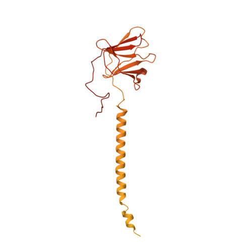
6OEG

Structure of CagX from a cryo-EM reconstruction of a T4SS


Experimental Data Snapshot

  • Method: ELECTRON MICROSCOPY
  • Resolution: 3.80 Å
  • Aggregation State: PARTICLE 
  • Reconstruction Method: SINGLE PARTICLE 

wwPDB Validation   3D Report Full Report


This is version 1.3 of the entry. See complete history


Literature

Structure of the Helicobacter pylori Cag type IV secretion system.

Chung, J.M.Sheedlo, M.J.Campbell, A.M.Sawhney, N.Frick-Cheng, A.E.Lacy, D.B.Cover, T.L.Ohi, M.D.

(2019) Elife 8

  • DOI: https://doi.org/10.7554/eLife.47644
  • Primary Citation of Related Structures:  
    6ODI, 6ODJ, 6OEE, 6OEF, 6OEG, 6OEH

  • PubMed Abstract: 

    Bacterial type IV secretion systems (T4SSs) are molecular machines that can mediate interbacterial DNA transfer through conjugation and delivery of effector molecules into host cells. The Helicobacter pylori Cag T4SS translocates CagA, a bacterial oncoprotein, into gastric cells, contributing to gastric cancer pathogenesis. We report the structure of a membrane-spanning Cag T4SS assembly, which we describe as three sub-assemblies: a 14-fold symmetric outer membrane core complex (OMCC), 17-fold symmetric periplasmic ring complex (PRC), and central stalk. Features that differ markedly from those of prototypical T4SSs include an expanded OMCC and unexpected symmetry mismatch between the OMCC and PRC. This structure is one of the largest bacterial secretion system assemblies ever reported and illustrates the remarkable structural diversity that exists among bacterial T4SSs.


  • Organizational Affiliation

    Life Sciences Institute, University of Michigan, Ann Arbor, United States.


Macromolecules
Find similar proteins by:  (by identity cutoff)  |  3D Structure
Entity ID: 1
MoleculeChains Sequence LengthOrganismDetailsImage
Type IV secretion system apparatus protein CagX522Helicobacter pyloriMutation(s): 0 
Entity Groups  
Sequence Clusters30% Identity50% Identity70% Identity90% Identity95% Identity100% Identity
Sequence Annotations
Expand
  • Reference Sequence
Experimental Data & Validation

Experimental Data

  • Method: ELECTRON MICROSCOPY
  • Resolution: 3.80 Å
  • Aggregation State: PARTICLE 
  • Reconstruction Method: SINGLE PARTICLE 

Structure Validation

View Full Validation Report


Entry History & Funding Information

Deposition Data


Funding OrganizationLocationGrant Number
National Institutes of Health/National Institute Of Allergy and Infectious Diseases (NIH/NIAID)United StatesNIH AI118932
National Institutes of Health/National Cancer Institute (NIH/NCI)United StatesCA116087
National Institutes of Health/National Institute of General Medical Sciences (NIH/NIGMS)United StatesGM103310
Other governmentUnited States1I01BX004447

Revision History  (Full details and data files)

  • Version 1.0: 2019-07-03
    Type: Initial release
  • Version 1.1: 2019-12-04
    Changes: Author supporting evidence
  • Version 1.2: 2020-10-07
    Changes: Database references
  • Version 1.3: 2024-03-13
    Changes: Data collection, Database references