6OBN

The crystal structure of coexpressed SDS22:PP1 complex


Experimental Data Snapshot

  • Method: X-RAY DIFFRACTION
  • Resolution: 2.70 Å
  • R-Value Free: 0.246 
  • R-Value Work: 0.202 
  • R-Value Observed: 0.204 

wwPDB Validation   3D Report Full Report


This is version 1.4 of the entry. See complete history


Literature

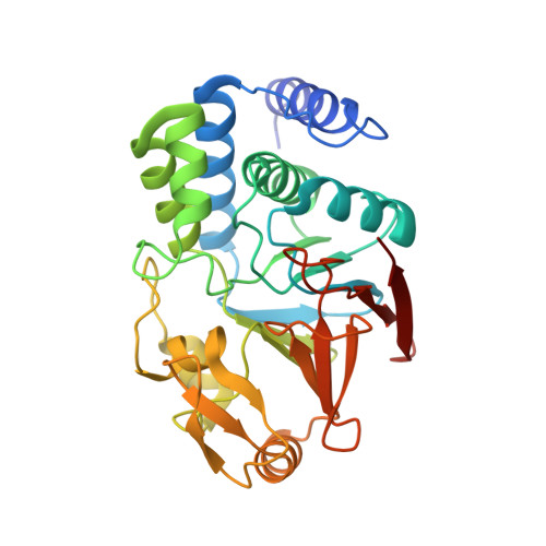
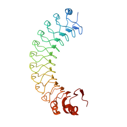
SDS22 selectively recognizes and traps metal-deficient inactive PP1.

Choy, M.S.Moon, T.M.Ravindran, R.Bray, J.A.Robinson, L.C.Archuleta, T.L.Shi, W.Peti, W.Tatchell, K.Page, R.

(2019) Proc Natl Acad Sci U S A 116: 20472-20481

  • DOI: https://doi.org/10.1073/pnas.1908718116
  • Primary Citation of Related Structures:  
    6OBN, 6OBP, 6OBQ, 6OBR, 6OBS, 6OBU

  • PubMed Abstract: 
  • The metalloenzyme protein phosphatase 1 (PP1), which is responsible for ≥50% of all dephosphorylation reactions, is regulated by scores of regulatory proteins, including the highly conserved SDS22 protein. SDS22 has numerous diverse functions, surprisingly acting as both a PP1 inhibitor and as an activator ...

    The metalloenzyme protein phosphatase 1 (PP1), which is responsible for ≥50% of all dephosphorylation reactions, is regulated by scores of regulatory proteins, including the highly conserved SDS22 protein. SDS22 has numerous diverse functions, surprisingly acting as both a PP1 inhibitor and as an activator. Here, we integrate cellular, biophysical, and crystallographic studies to address this conundrum. We discovered that SDS22 selectively binds a unique conformation of PP1 that contains a single metal (M2) at its active site, i.e., SDS22 traps metal-deficient inactive PP1. Furthermore, we showed that SDS22 dissociation is accompanied by a second metal (M1) being loaded into PP1, as free metal cannot dissociate the complex and M1-deficient mutants remain constitutively trapped by SDS22. Together, our findings reveal that M1 metal loading and loss are essential for PP1 regulation in cells, which has broad implications for PP1 maturation, activity, and holoenzyme subunit exchange.


    Organizational Affiliation

    Department of Chemistry and Biochemistry, University of Arizona, Tucson, AZ 85721; ktatch@lsuhsc.edu rebeccapage@email.arizona.edu.


Macromolecules
Find similar proteins by:  (by identity cutoff)  |  3D Structure
Entity ID: 1
MoleculeChainsSequence LengthOrganismDetailsImage
Serine/threonine-protein phosphatase PP1-alpha catalytic subunitA, B305Homo sapiensMutation(s): 0 
Gene Names: PPP1CAPPP1A
EC: 3.1.3.16
UniProt & NIH Common Fund Data Resources
Find proteins for P62136 (Homo sapiens)
Explore P62136 
Go to UniProtKB:  P62136
PHAROS:  P62136
Entity Groups  
Sequence Clusters30% Identity50% Identity70% Identity90% Identity95% Identity100% Identity
UniProt GroupP62136
Protein Feature View
Expand
  • Reference Sequence
Find similar proteins by:  (by identity cutoff)  |  3D Structure
Entity ID: 2
MoleculeChainsSequence LengthOrganismDetailsImage
Protein phosphatase 1 regulatory subunit 7C, D310Homo sapiensMutation(s): 0 
Gene Names: PPP1R7SDS22
UniProt & NIH Common Fund Data Resources
Find proteins for Q15435 (Homo sapiens)
Explore Q15435 
Go to UniProtKB:  Q15435
PHAROS:  Q15435
Entity Groups  
Sequence Clusters30% Identity50% Identity70% Identity90% Identity95% Identity100% Identity
UniProt GroupQ15435
Protein Feature View
Expand
  • Reference Sequence
Small Molecules
Ligands 4 Unique
IDChainsName / Formula / InChI Key2D Diagram3D Interactions
MPD
Query on MPD

Download Ideal Coordinates CCD File 
J [auth A],
K [auth A],
R [auth C],
S [auth C]
(4S)-2-METHYL-2,4-PENTANEDIOL
C6 H14 O2
SVTBMSDMJJWYQN-YFKPBYRVSA-N
 Ligand Interaction
PO4
Query on PO4

Download Ideal Coordinates CCD File 
E [auth A],
F [auth A],
L [auth B],
O [auth C],
P [auth C],
E [auth A],
F [auth A],
L [auth B],
O [auth C],
P [auth C],
Q [auth C],
T [auth D],
U [auth D]
PHOSPHATE ION
O4 P
NBIIXXVUZAFLBC-UHFFFAOYSA-K
 Ligand Interaction
FE
Query on FE

Download Ideal Coordinates CCD File 
G [auth A],
M [auth B]
FE (III) ION
Fe
VTLYFUHAOXGGBS-UHFFFAOYSA-N
 Ligand Interaction
CL
Query on CL

Download Ideal Coordinates CCD File 
H [auth A],
I [auth A],
N [auth B]
CHLORIDE ION
Cl
VEXZGXHMUGYJMC-UHFFFAOYSA-M
 Ligand Interaction
Experimental Data & Validation

Experimental Data

  • Method: X-RAY DIFFRACTION
  • Resolution: 2.70 Å
  • R-Value Free: 0.246 
  • R-Value Work: 0.202 
  • R-Value Observed: 0.204 
  • Space Group: P 65 2 2
Unit Cell:
Length ( Å )Angle ( ˚ )
a = 94.147α = 90
b = 94.147β = 90
c = 545.819γ = 120
Software Package:
Software NamePurpose
PHENIXrefinement
XDSdata reduction
Aimlessdata scaling
PHENIXphasing

Structure Validation

View Full Validation Report


Entry History & Funding Information

Deposition Data


Funding OrganizationLocationGrant Number
National Institutes of Health/National Human Genome Research Institute (NIH/NHGRI)United States--

Revision History  (Full details and data files)

  • Version 1.0: 2019-09-18
    Type: Initial release
  • Version 1.1: 2019-09-25
    Changes: Data collection, Database references
  • Version 1.2: 2019-10-02
    Changes: Data collection, Database references
  • Version 1.3: 2019-10-23
    Changes: Data collection, Database references
  • Version 1.4: 2019-12-18
    Changes: Author supporting evidence