6NYA

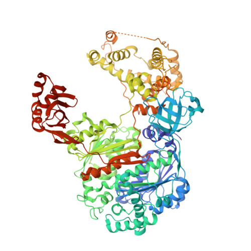
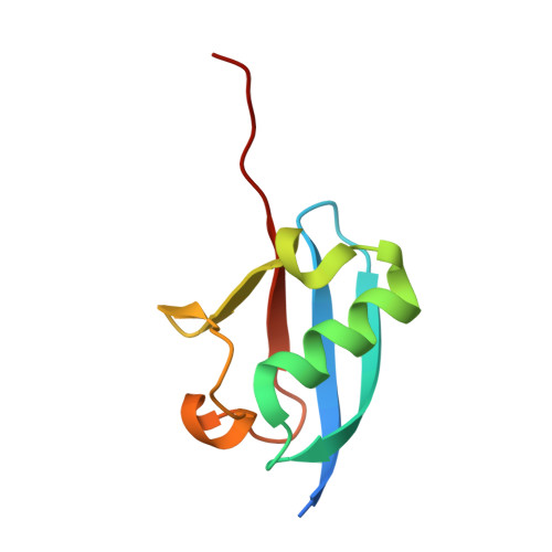
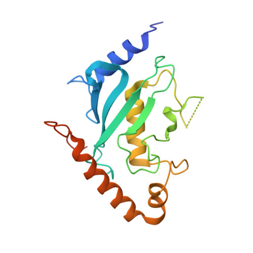
Crystal Structure of ubiquitin E1 (Uba1) in complex with Ubc3 (Cdc34) and ubiquitin


Experimental Data Snapshot

  • Method: X-RAY DIFFRACTION
  • Resolution: 2.06 Å
  • R-Value Free: 0.217 
  • R-Value Work: 0.187 
  • R-Value Observed: 0.188 

Starting Model: experimental
View more details

wwPDB Validation   3D Report Full Report


Ligand Structure Quality Assessment 


This is version 1.2 of the entry. See complete history


Literature

Structural insights into E1 recognition and the ubiquitin-conjugating activity of the E2 enzyme Cdc34.

Williams, K.M.Qie, S.Atkison, J.H.Salazar-Arango, S.Alan Diehl, J.Olsen, S.K.

(2019) Nat Commun 10: 3296-3296

  • DOI: https://doi.org/10.1038/s41467-019-11061-8
  • Primary Citation of Related Structures:  
    6NYA, 6NYD, 6NYO

  • PubMed Abstract: 

    Ubiquitin (Ub) signaling requires the sequential interactions and activities of three enzymes, E1, E2, and E3. Cdc34 is an E2 that plays a key role in regulating cell cycle progression and requires unique structural elements to function. The molecular basis by which Cdc34 engages its E1 and the structural mechanisms by which its unique C-terminal extension functions in Cdc34 activity are unknown. Here, we present crystal structures of Cdc34 alone and in complex with E1, and a Cdc34~Ub thioester mimetic that represents the product of Uba1-Cdc34 Ub transthiolation. These structures reveal conformational changes in Uba1 and Cdc34 and a unique binding mode that are required for transthiolation. The Cdc34~Ub structure reveals contacts between the Cdc34 C-terminal extension and Ub that stabilize Cdc34~Ub in a closed conformation and are critical for Ub discharge. Altogether, our structural, biochemical, and cell-based studies provide insights into the molecular mechanisms by which Cdc34 function in cells.


  • Organizational Affiliation

    Department of Biochemistry & Molecular Biology and Hollings Cancer Center, Medical University of South Carolina, Charleston, SC, 29425, USA.


Macromolecules
Find similar proteins by:  (by identity cutoff)  |  3D Structure
Entity ID: 1
MoleculeChains Sequence LengthOrganismDetailsImage
Ubiquitin-activating enzyme E1 1
A, D
1,017Saccharomyces cerevisiae S288CMutation(s): 0 
Gene Names: UBA1YKL210W
EC: 6.2.1.45
UniProt
Find proteins for P22515 (Saccharomyces cerevisiae (strain ATCC 204508 / S288c))
Explore P22515 
Go to UniProtKB:  P22515
Entity Groups  
Sequence Clusters30% Identity50% Identity70% Identity90% Identity95% Identity100% Identity
UniProt GroupP22515
Sequence Annotations
Expand
  • Reference Sequence
Find similar proteins by:  (by identity cutoff)  |  3D Structure
Entity ID: 2
MoleculeChains Sequence LengthOrganismDetailsImage
Ubiquitin
B, E
85Triticum aestivumMutation(s): 7 
UniProt
Find proteins for P69326 (Triticum aestivum)
Explore P69326 
Go to UniProtKB:  P69326
Entity Groups  
Sequence Clusters30% Identity50% Identity70% Identity90% Identity95% Identity100% Identity
UniProt GroupP69326
Sequence Annotations
Expand
  • Reference Sequence
Find similar proteins by:  (by identity cutoff)  |  3D Structure
Entity ID: 3
MoleculeChains Sequence LengthOrganismDetailsImage
Ubiquitin-conjugating enzyme E2-34 kDa
C, F
197Saccharomyces cerevisiae S288CMutation(s): 0 
Gene Names: CDC34DNA6UBC3YDR054CD4211YD9609.08C
EC: 2.3.2.23
UniProt
Find proteins for P14682 (Saccharomyces cerevisiae (strain ATCC 204508 / S288c))
Explore P14682 
Go to UniProtKB:  P14682
Entity Groups  
Sequence Clusters30% Identity50% Identity70% Identity90% Identity95% Identity100% Identity
UniProt GroupP14682
Sequence Annotations
Expand
  • Reference Sequence
Small Molecules
Ligands 4 Unique
IDChains Name / Formula / InChI Key2D Diagram3D Interactions
ATP
Query on ATP

Download Ideal Coordinates CCD File 
DA [auth D],
G [auth A]
ADENOSINE-5'-TRIPHOSPHATE
C10 H16 N5 O13 P3
ZKHQWZAMYRWXGA-KQYNXXCUSA-N
SO4
Query on SO4

Download Ideal Coordinates CCD File 
EA [auth D]
FA [auth D]
GA [auth D]
HA [auth D]
I [auth A]
EA [auth D],
FA [auth D],
GA [auth D],
HA [auth D],
I [auth A],
IA [auth D],
J [auth A],
JA [auth D],
K [auth A],
KA [auth D],
L [auth A],
M [auth A],
N [auth A],
O [auth A],
P [auth A]
SULFATE ION
O4 S
QAOWNCQODCNURD-UHFFFAOYSA-L
EDO
Query on EDO

Download Ideal Coordinates CCD File 
AA [auth A]
BA [auth C]
LA [auth D]
MA [auth D]
NA [auth D]
AA [auth A],
BA [auth C],
LA [auth D],
MA [auth D],
NA [auth D],
OA [auth D],
PA [auth D],
Q [auth A],
QA [auth D],
R [auth A],
RA [auth D],
S [auth A],
SA [auth D],
T [auth A],
TA [auth D],
U [auth A],
V [auth A],
W [auth A],
X [auth A],
Y [auth A],
Z [auth A]
1,2-ETHANEDIOL
C2 H6 O2
LYCAIKOWRPUZTN-UHFFFAOYSA-N
MG
Query on MG

Download Ideal Coordinates CCD File 
CA [auth D],
H [auth A]
MAGNESIUM ION
Mg
JLVVSXFLKOJNIY-UHFFFAOYSA-N
Experimental Data & Validation

Experimental Data

  • Method: X-RAY DIFFRACTION
  • Resolution: 2.06 Å
  • R-Value Free: 0.217 
  • R-Value Work: 0.187 
  • R-Value Observed: 0.188 
  • Space Group: P 1 21 1
Unit Cell:
Length ( Å )Angle ( ˚ )
a = 126.445α = 90
b = 68.544β = 110.72
c = 171.721γ = 90
Software Package:
Software NamePurpose
PHENIXrefinement
XDSdata reduction
Aimlessdata scaling
PHASERphasing

Structure Validation

View Full Validation Report


Ligand Structure Quality Assessment 


Entry History & Funding Information

Deposition Data


Funding OrganizationLocationGrant Number
National Institutes of Health/National Institute of General Medical Sciences (NIH/NIGMS)United StatesGM115568

Revision History  (Full details and data files)

  • Version 1.0: 2019-08-07
    Type: Initial release
  • Version 1.1: 2020-01-01
    Changes: Author supporting evidence
  • Version 1.2: 2023-10-11
    Changes: Data collection, Database references, Derived calculations, Refinement description