Crystal Structure of d-Ornithine/d-Lysine Decarboxylase, a Stereoinverting Decarboxylase: Implications for Substrate Specificity and Stereospecificity of Fold III Decarboxylases.
Phillips, R.S., Poteh, P., Krajcovic, D., Miller, K.A., Hoover, T.R.(2019) Biochemistry 58: 1038-1042
- PubMed: 30699288 Search on PubMed
- DOI: https://doi.org/10.1021/acs.biochem.8b01319
- Primary Citation Related Structures:
6N2H - PubMed Abstract:
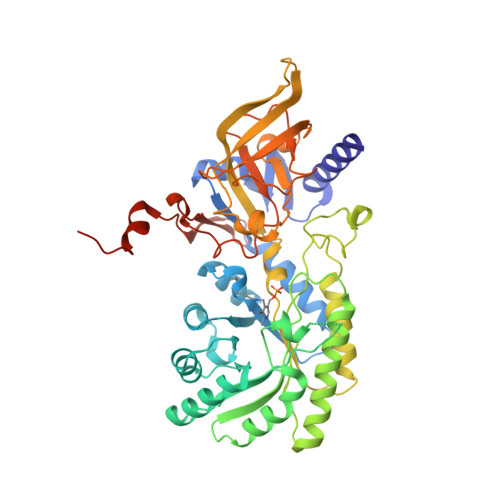
A newly discovered Fold III pyridoxal 5'-phosphate (PLP)-dependent decarboxylase, d-ornithine/lysine decarboxylase (DOKDC), catalyzes decarboxylation of d-lysine and d-ornithine with inversion of stereochemistry. The X-ray crystal structure of DOKDC has been determined to 1.72 Å. DOKDC has a low level of sequence identity (<30%) with meso-diaminopimelate decarboxylase (DAPDC) and l-lysine/ornithine decarboxylase (LODC), but its three-dimensional structure is very similar. The distal binding site of DAPDC contains a conserved arginine that forms an ion pair with the l-carboxylate end of DAP. In both LODC and DOKDC, this distal site is modified by replacement of the arginine with aspartate, changing the substrate specificity. l-Ornithine decarboxylase (ODC) and LODC have a conserved phenylalanine on the re-face of the PLP complex that has been found to play a key role in the decarboxylation mechanism. We have found that both DAPDC and DOKDC have tyrosine instead of phenylalanine at this position, which precludes the binding of l-amino acids. Because the PLP-binding lysine in ODC, LODC, DAPDC, and DOKDC is located on the re-face of the PLP, we propose that this is the acid group responsible for protonation of the product, thus resulting in the observed retention of configuration for decarboxylation of l-amino acids and inversion for decarboxylation of d-amino acids. The reactions of DAPDC and DOKDC are likely accelerated by positive electrostatics on the re-face by the lysine ε-ammonium ion and on the si-face by closure of the lid over the active site, resulting in desolvation and destabilization of the d-amino acid carboxylate.
- Department of Chemistry , University of Georgia , Athens , Georgia 30602 , United States.
Organizational Affiliation: