Evidence for distinct rate-limiting steps in the cleavage of alkenes by carotenoid cleavage dioxygenases.
Khadka, N., Farquhar, E.R., Hill, H.E., Shi, W., von Lintig, J., Kiser, P.D.(2019) J Biological Chem 294: 10596-10606
- PubMed: 31138651 Search on PubMedSearch on PubMed Central
- DOI: https://doi.org/10.1074/jbc.RA119.007535
- Primary Citation Related Structures:
6N1Y, 6N20, 6N21 - PubMed Abstract:
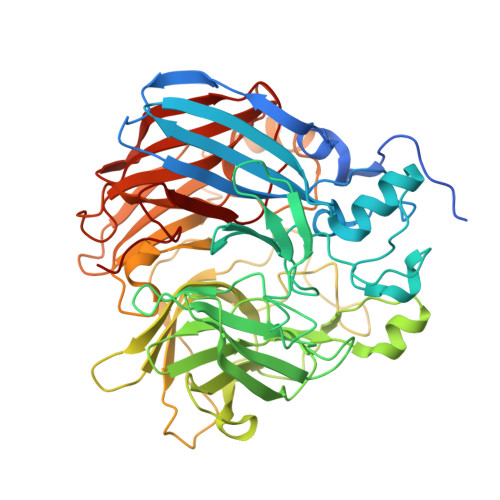
Carotenoid cleavage dioxygenases (CCDs) use a nonheme Fe(II) cofactor to split alkene bonds of carotenoid and stilbenoid substrates. The iron centers of CCDs are typically five-coordinate in their resting states, with solvent occupying an exchangeable site. The involvement of this iron-bound solvent in CCD catalysis has not been experimentally addressed, but computational studies suggest two possible roles. 1) Solvent dissociation provides a coordination site for O 2 , or 2) solvent remains bound to iron but changes its equilibrium position to allow O 2 binding and potentially acts as a proton source. To test these predictions, we investigated isotope effects (H 2 O versus D 2 O) on two stilbenoid-cleaving CCDs, Novosphingobium aromaticivorans oxygenase 2 (NOV2) and Neurospora crassa carotenoid oxygenase 1 (CAO1), using piceatannol as a substrate. NOV2 exhibited an inverse isotope effect ( k H / k D ∼ 0.6) in an air-saturated buffer, suggesting that solvent dissociates from iron during the catalytic cycle. By contrast, CAO1 displayed a normal isotope effect ( k H / k D ∼ 1.7), suggesting proton transfer in the rate-limiting step. X-ray absorption spectroscopy on NOV2 and CAO1 indicated that the protonation states of the iron ligands are unchanged within pH 6.5-8.5 and that the Fe(II)-aquo bond is minimally altered by substrate binding. We pinpointed the origin of the differential kinetic behaviors of NOV2 and CAO1 to a single amino acid difference near the solvent-binding site of iron, and X-ray crystallography revealed that the substitution alters binding of diffusible ligands to the iron center. We conclude that solvent-iron dissociation and proton transfer are both associated with the CCD catalytic mechanism.
- From the Department of Pharmacology, School of Medicine, Case Western Reserve University, Cleveland, Ohio 44106.
Organizational Affiliation: