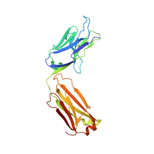
6MY5

Crystal structure of the dimeric bH1-Fab variant [HC-Y33W,HC-D98F,HC-G99M,LC-S30bR]

  • Classification: IMMUNE SYSTEM
  • Organism(s): Homo sapiens
  • Expression System: Cricetulus griseus
  • Mutation(s): No 

  • Deposited: 2018-11-01 Released: 2019-07-31 
  • Deposition Author(s): Shi, R.
  • Funding Organization(s): Natural Sciences and Engineering Research Council (NSERC, Canada)

Experimental Data Snapshot

  • Method: X-RAY DIFFRACTION
  • Resolution: 1.73 Å
  • R-Value Free: 0.229 
  • R-Value Work: 0.186 
  • R-Value Observed: 0.188 

Starting Model: experimental
View more details

wwPDB Validation   3D Report Full Report


This is version 1.4 of the entry. See complete history


Literature

Binding symmetry and surface flexibility mediate antibody self-association.

Schrag, J.D.Picard, M.E.Gaudreault, F.Gagnon, L.P.Baardsnes, J.Manenda, M.S.Sheff, J.Deprez, C.Baptista, C.Hogues, H.Kelly, J.F.Purisima, E.O.Shi, R.Sulea, T.

(2019) MAbs 11: 1300-1318

  • DOI: https://doi.org/10.1080/19420862.2019.1632114
  • Primary Citation of Related Structures:  
    6MXR, 6MXS, 6MY4, 6MY5

  • PubMed Abstract: 

    Solution stability is an important factor in the optimization of engineered biotherapeutic candidates such as monoclonal antibodies because of its possible effects on manufacturability, pharmacology, efficacy and safety. A detailed atomic understanding of the mechanisms governing self-association of natively folded protein monomers is required to devise predictive tools to guide screening and re-engineering along the drug development pipeline. We investigated pairs of affinity-matured full-size antibodies and observed drastically different propensities to aggregate from variants differing by a single amino-acid. Biophysical testing showed that antigen-binding fragments (Fabs) from the aggregating antibodies also reversibly associated with equilibrium dissociation constants in the low-micromolar range. Crystal structures (PDB accession codes 6MXR, 6MXS, 6MY4, 6MY5) and bottom-up hydrogen-exchange mass spectrometry revealed that Fab self-association occurs in a symmetric mode that involves the antigen complementarity-determining regions. Subtle local conformational changes incurred upon point mutation of monomeric variants foster formation of complementary polar interactions and hydrophobic contacts to generate a dimeric Fab interface. Testing of popular in silico tools generally indicated low reliabilities for predicting the aggregation propensities observed. A structure-aggregation data set is provided here in order to stimulate further improvements of in silico tools for prediction of native aggregation. Incorporation of intermolecular docking, conformational flexibility, and short-range packing interactions may all be necessary features of the ideal algorithm.


  • Organizational Affiliation

    Human Health Therapeutics Research Centre, National Research Council Canada , Montreal , QC H4P 2R2 , Canada.


Macromolecules
Find similar proteins by:  (by identity cutoff)  |  3D Structure
Entity ID: 1
MoleculeChains Sequence LengthOrganismDetailsImage
anti-VEGF-A Fab fragment bH1 heavy chainA [auth H],
C [auth A]
236Homo sapiensMutation(s): 0 
UniProt
Find proteins for V9HW68 (Homo sapiens)
Explore V9HW68 
Go to UniProtKB:  V9HW68
Entity Groups  
Sequence Clusters30% Identity50% Identity70% Identity90% Identity95% Identity100% Identity
UniProt GroupV9HW68
Sequence Annotations
Expand
  • Reference Sequence
Find similar proteins by:  (by identity cutoff)  |  3D Structure
Entity ID: 2
MoleculeChains Sequence LengthOrganismDetailsImage
anti-VEGF-A Fab fragment bH1 light chainB [auth L],
D [auth B]
218Homo sapiensMutation(s): 0 
UniProt
Find proteins for Q7Z3Y4 (Homo sapiens)
Explore Q7Z3Y4 
Go to UniProtKB:  Q7Z3Y4
Entity Groups  
Sequence Clusters30% Identity50% Identity70% Identity90% Identity95% Identity100% Identity
UniProt GroupQ7Z3Y4
Sequence Annotations
Expand
  • Reference Sequence
Experimental Data & Validation

Experimental Data

  • Method: X-RAY DIFFRACTION
  • Resolution: 1.73 Å
  • R-Value Free: 0.229 
  • R-Value Work: 0.186 
  • R-Value Observed: 0.188 
  • Space Group: P 1 21 1
Unit Cell:
Length ( Å )Angle ( ˚ )
a = 81.599α = 90
b = 65.53β = 100.73
c = 92.56γ = 90
Software Package:
Software NamePurpose
REFMACrefinement
Aimlessdata scaling
PDB_EXTRACTdata extraction
iMOSFLMdata reduction
MOLREPphasing

Structure Validation

View Full Validation Report


Entry History & Funding Information

Deposition Data

  • Released Date: 2019-07-31 
  • Deposition Author(s): Shi, R.

Funding OrganizationLocationGrant Number
Natural Sciences and Engineering Research Council (NSERC, Canada)Canada436202

Revision History  (Full details and data files)

  • Version 1.0: 2019-07-31
    Type: Initial release
  • Version 1.1: 2019-09-25
    Changes: Data collection, Database references
  • Version 1.2: 2020-01-08
    Changes: Author supporting evidence
  • Version 1.3: 2023-10-11
    Changes: Data collection, Database references, Refinement description
  • Version 1.4: 2024-11-20
    Changes: Structure summary