6MS3

Crystal structure of the GH43 protein BlXynB mutant (K247S) from Bacillus licheniformis


Experimental Data Snapshot

  • Method: X-RAY DIFFRACTION
  • Resolution: 1.95 Å
  • R-Value Free: 0.201 
  • R-Value Work: 0.168 
  • R-Value Observed: 0.168 

wwPDB Validation   3D Report Full Report


This is version 1.2 of the entry. See complete history


Literature

Structure-guided design combined with evolutionary diversity led to the discovery of the xylose-releasing exo-xylanase activity in the glycoside hydrolase family 43.

Zanphorlin, L.M.de Morais, M.A.B.Diogo, J.A.Domingues, M.N.de Souza, F.H.M.Ruller, R.Murakami, M.T.

(2019) Biotechnol Bioeng 116: 734-744

  • DOI: https://doi.org/10.1002/bit.26899
  • Primary Citation of Related Structures:  
    6MS2, 6MS3

  • PubMed Abstract: 

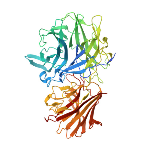
    Rational design is an important tool for sculpting functional and stability properties of proteins and its potential can be much magnified when combined with in vitro and natural evolutionary diversity. Herein, we report the structure-guided design of a xylose-releasing exo-β-1,4-xylanase from an inactive member of glycoside hydrolase family 43 (GH43). Structural analysis revealed a nonconserved substitution (Lys 247 ) that results in the disruption of the hydrogen bond network that supports catalysis. The mutation of this residue to a conserved serine restored the catalytic activity and crystal structure elucidation of the mutant confirmed the recovery of the proper orientation of the catalytically relevant histidine. Interestingly, the tailored enzyme can cleave both xylooligosaccharides and xylan, releasing xylose as the main product, being the first xylose-releasing exo-β-1,4-xylanase reported in the GH43 family. This enzyme presents a unique active-site topology when compared with closely related β-xylosidases, which is the absence of a hydrophobic barrier at the positive-subsite region, allowing the accommodation of long substrates. Therefore, the combination of rational design for catalytic activation along with naturally occurring differences in the substrate binding interface led to the discovery of a novel activity within the GH43 family. In addition, these results demonstrate the importance of solvation of the β-propeller hollow for GH43 catalytic function and expand our mechanistic understanding about the diverse modes of action of GH43 members, a key and polyspecific carbohydrate-active enzyme family abundant in most plant cell-wall-degrading microorganisms.


  • Organizational Affiliation

    Brazilian Bioethanol Science and Technology Laboratory, National Center for Research in Energy and Materials, Campinas, São Paulo, Brazil.


Macromolecules
Find similar proteins by:  (by identity cutoff)  |  3D Structure
Entity ID: 1
MoleculeChains Sequence LengthOrganismDetailsImage
Glycoside Hydrolase Family 43
A, B
538Bacillus licheniformis DSM 13 = ATCC 14580Mutation(s): 1 
Gene Names: xynBBL03023
UniProt
Find proteins for Q65MB6 (Bacillus licheniformis (strain ATCC 14580 / DSM 13 / JCM 2505 / CCUG 7422 / NBRC 12200 / NCIMB 9375 / NCTC 10341 / NRRL NRS-1264 / Gibson 46))
Explore Q65MB6 
Go to UniProtKB:  Q65MB6
Entity Groups  
Sequence Clusters30% Identity50% Identity70% Identity90% Identity95% Identity100% Identity
UniProt GroupQ65MB6
Sequence Annotations
Expand
  • Reference Sequence
Small Molecules
Ligands 4 Unique
IDChains Name / Formula / InChI Key2D Diagram3D Interactions
SO4
Query on SO4

Download Ideal Coordinates CCD File 
D [auth A]
E [auth A]
F [auth A]
G [auth A]
H [auth A]
D [auth A],
E [auth A],
F [auth A],
G [auth A],
H [auth A],
L [auth B],
M [auth B],
N [auth B],
O [auth B],
P [auth B],
Q [auth B]
SULFATE ION
O4 S
QAOWNCQODCNURD-UHFFFAOYSA-L
GOL
Query on GOL

Download Ideal Coordinates CCD File 
I [auth A],
R [auth B]
GLYCEROL
C3 H8 O3
PEDCQBHIVMGVHV-UHFFFAOYSA-N
IMD
Query on IMD

Download Ideal Coordinates CCD File 
J [auth A],
S [auth B]
IMIDAZOLE
C3 H5 N2
RAXXELZNTBOGNW-UHFFFAOYSA-O
CA
Query on CA

Download Ideal Coordinates CCD File 
C [auth A],
K [auth B]
CALCIUM ION
Ca
BHPQYMZQTOCNFJ-UHFFFAOYSA-N
Experimental Data & Validation

Experimental Data

  • Method: X-RAY DIFFRACTION
  • Resolution: 1.95 Å
  • R-Value Free: 0.201 
  • R-Value Work: 0.168 
  • R-Value Observed: 0.168 
  • Space Group: P 43
Unit Cell:
Length ( Å )Angle ( ˚ )
a = 82.126α = 90
b = 82.126β = 90
c = 173.236γ = 90
Software Package:
Software NamePurpose
XSCALEdata scaling
PHENIXrefinement
PDB_EXTRACTdata extraction
XDSdata reduction
PHASERphasing

Structure Validation

View Full Validation Report


Entry History & Funding Information

Deposition Data


Funding OrganizationLocationGrant Number
Sao Paulo Research Foundation (FAPESP)Brazil--

Revision History  (Full details and data files)

  • Version 1.0: 2019-04-17
    Type: Initial release
  • Version 1.1: 2020-01-01
    Changes: Author supporting evidence
  • Version 1.2: 2024-03-13
    Changes: Data collection, Database references, Derived calculations