6MOZ | pdb_00006moz

Structure of acid-beta-glucosidase in complex with an aromatic pyrrolidine iminosugar inhibitor


Experimental Data Snapshot

  • Method: X-RAY DIFFRACTION
  • Resolution: 2.10 Å
  • R-Value Free: 
    0.228 (Depositor), 0.226 (DCC) 
  • R-Value Work: 
    0.200 (Depositor) 
  • R-Value Observed: 
    0.201 (Depositor) 

Starting Model: experimental
View more details

wwPDB Validation 3D Report Full Report

Validation slider image for 6MOZ

Ligand Structure Quality Assessment 


This is version 1.4 of the entry. See complete history

Literature

Exploring substituent diversity on pyrrolidine-aryltriazole iminosugars: Structural basis of beta-glucocerebrosidase inhibition.

Martinez-Bailen, M.Carmona, A.T.Patterson-Orazem, A.C.Lieberman, R.L.Ide, D.Kubo, M.Kato, A.Robina, I.Moreno-Vargas, A.J.

(2019) Bioorg Chem 86: 652-664

  • DOI: https://doi.org/10.1016/j.bioorg.2019.02.025
  • Primary Citation Related Structures: 
    6MOZ

  • PubMed Abstract: 

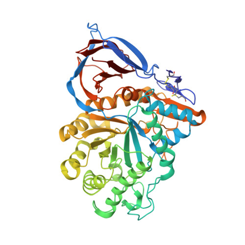
    The synthesis of a library of pyrrolidine-aryltriazole hybrids through CuAAC between two epimeric dihydroxylated azidomethylpyrrolidines and differently substituted phenylacetylenes is reported. The evaluation of the new compounds as inhibitors of lysosomal β-glucocerebrosidase showed the importance of the substitution pattern of the phenyl moiety in the inhibition. Crystallization and docking studies revealed key interactions of the pyrrolidine motif with aminoacid residues of the catalytic site while the aryltriazole moiety extended along a hydrophobic surface groove. Some of these compounds were able to increase the enzyme activity in Gaucher patient fibroblasts, acting as a new type of chemical chaperone for Gaucher disease.


  • Organizational Affiliation
    • Department of Organic Chemistry, Faculty of Chemistry, University of Seville, C/Prof. García González, 1, 41012-Seville, Spain.

Macromolecule Content 

  • Total Structure Weight: 113.45 kDa 
  • Atom Count: 8,118 
  • Modeled Residue Count: 994 
  • Deposited Residue Count: 994 
  • Unique protein chains: 1

Macromolecules

Find similar proteins by:|  3D Structure
Entity ID: 1
MoleculeChains  Sequence LengthOrganismDetailsImage
Glucosylceramidase
A, B
497Homo sapiensMutation(s): 1 
Gene Names: GBAGCGLUC
EC: 3.2.1.45 (PDB Primary Data), 3.2.1.46 (UniProt), 2.4.1 (UniProt), 3.2.1 (UniProt)
UniProt & NIH Common Fund Data Resources
Find proteins for P04062 (Homo sapiens)
Explore P04062 
Go to UniProtKB:  P04062
PHAROS:  P04062
GTEx:  ENSG00000177628 
Entity Groups
Sequence Clusters30% Identity50% Identity70% Identity90% Identity95% Identity100% Identity
UniProt GroupP04062
Glycosylation
Glycosylation Sites: 1Go to GlyGen: P04062-1
Sequence Annotations
Expand
Reference Sequence

Small Molecules

Ligands 5 Unique
IDChains Name / Formula / InChI Key2D Diagram3D Interactions
JXA
(Subject of Investigation/LOI)

Query on JXA


Download:Ideal Coordinates CCD File
S [auth A](2R,3S,4R)-2-{[4-(3,5-dichlorophenyl)-1H-1,2,3-triazol-1-yl]methyl}pyrrolidine-3,4-diol
C13 H14 Cl2 N4 O2
QVWPLFTVMWIDFF-UPJWGTAASA-N
NAG

Query on NAG


Download:Ideal Coordinates CCD File
C [auth A],
W [auth B]
2-acetamido-2-deoxy-beta-D-glucopyranose
C8 H15 N O6
OVRNDRQMDRJTHS-FMDGEEDCSA-N
PO4

Query on PO4


Download:Ideal Coordinates CCD File
PA [auth B]
QA [auth B]
RA [auth B]
T [auth A]
U [auth A]
PA [auth B],
QA [auth B],
RA [auth B],
T [auth A],
U [auth A],
V [auth A]
PHOSPHATE ION
O4 P
NBIIXXVUZAFLBC-UHFFFAOYSA-K
GOL

Query on GOL


Download:Ideal Coordinates CCD File
OA [auth B]GLYCEROL
C3 H8 O3
PEDCQBHIVMGVHV-UHFFFAOYSA-N
NA

Query on NA


Download:Ideal Coordinates CCD File
AA [auth B]
BA [auth B]
CA [auth B]
D [auth A]
DA [auth B]
AA [auth B],
BA [auth B],
CA [auth B],
D [auth A],
DA [auth B],
E [auth A],
EA [auth B],
F [auth A],
FA [auth B],
G [auth A],
GA [auth B],
H [auth A],
HA [auth B],
I [auth A],
IA [auth B],
J [auth A],
JA [auth B],
K [auth A],
KA [auth B],
L [auth A],
LA [auth B],
M [auth A],
MA [auth B],
N [auth A],
NA [auth B],
O [auth A],
P [auth A],
Q [auth A],
R [auth A],
X [auth B],
Y [auth B],
Z [auth B]
SODIUM ION
Na
FKNQFGJONOIPTF-UHFFFAOYSA-N

Experimental Data & Validation

Experimental Data

  • Method: X-RAY DIFFRACTION
  • Resolution: 2.10 Å
  • R-Value Free:  0.228 (Depositor), 0.226 (DCC) 
  • R-Value Work:  0.200 (Depositor) 
  • R-Value Observed: 0.201 (Depositor) 
Space Group: C 2 2 21
Unit Cell:
Length ( Å )Angle ( ˚ )
a = 110.28α = 90
b = 285.01β = 90
c = 91.68γ = 90
Software Package:
Software NamePurpose
PHENIXrefinement
XDSdata reduction
XDSdata scaling
PHENIXphasing

Structure Validation

View Full Validation Report


Ligand Structure Quality Assessment 


Entry History 

& Funding Information

Deposition Data


Funding OrganizationLocationGrant Number
National Institutes of Health/National Eye Institute (NIH/NEI)United States021205

Revision History  (Full details and data files)

  • Version 1.0: 2019-06-19
    Type: Initial release
  • Version 1.1: 2019-12-11
    Changes: Author supporting evidence
  • Version 1.2: 2020-07-29
    Type: Remediation
    Reason: Carbohydrate remediation
    Changes: Data collection, Derived calculations, Structure summary
  • Version 1.3: 2023-10-11
    Changes: Data collection, Database references, Refinement description, Structure summary
  • Version 1.4: 2024-11-13
    Changes: Structure summary