6MMB | pdb_00006mmb

Diheteromeric NMDA receptor GluN1/GluN2A in the 'Super-Splayed' conformation, in complex with glycine and glutamate, in the presence of 1 micromolar zinc chloride, and at pH 6.1


Experimental Data Snapshot

  • Method: ELECTRON MICROSCOPY
  • Resolution: 12.7 Å
  • Aggregation State: PARTICLE 
  • Reconstruction Method: SINGLE PARTICLE 

Starting Models: experimental
View more details

wwPDB Validation 3D Report Full Report

Validation slider image for 6MMB

This is version 1.4 of the entry. See complete history

Literature

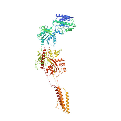
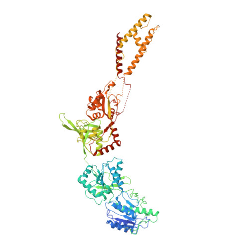
Mechanisms for Zinc and Proton Inhibition of the GluN1/GluN2A NMDA Receptor.

Jalali-Yazdi, F.Chowdhury, S.Yoshioka, C.Gouaux, E.

(2018) Cell 175: 1520-1532.e15

  • DOI: https://doi.org/10.1016/j.cell.2018.10.043
  • Primary Citation Related Structures: 
    6MM9, 6MMA, 6MMB, 6MMG, 6MMH, 6MMI, 6MMJ, 6MMK, 6MML, 6MMM, 6MMN, 6MMP, 6MMR, 6MMS, 6MMT, 6MMU, 6MMV, 6MMW, 6MMX

  • PubMed Abstract: 

    N-methyl-D-aspartate receptors (NMDARs) play essential roles in memory formation, neuronal plasticity, and brain development, with their dysfunction linked to a range of disorders from ischemia to schizophrenia. Zinc and pH are physiological allosteric modulators of NMDARs, with GluN2A-containing receptors inhibited by nanomolar concentrations of divalent zinc and by excursions to low pH. Despite the widespread importance of zinc and proton modulation of NMDARs, the molecular mechanism by which these ions modulate receptor activity has proven elusive. Here, we use cryoelectron microscopy to elucidate the structure of the GluN1/GluN2A NMDAR in a large ensemble of conformations under a range of physiologically relevant zinc and proton concentrations. We show how zinc binding to the amino terminal domain elicits structural changes that are transduced though the ligand-binding domain and result in constriction of the ion channel gate.


  • Organizational Affiliation
    • Vollum Institute, Oregon Health and Science University, Portland, OR 97239, USA.

Macromolecule Content 

  • Total Structure Weight: 383.38 kDa 
  • Atom Count: 24,494 
  • Modeled Residue Count: 3,039 
  • Deposited Residue Count: 3,350 
  • Unique protein chains: 2

Macromolecules

Find similar proteins by:|  3D Structure
Entity ID: 1
MoleculeChains  Sequence LengthOrganismDetailsImage
Glutamate receptor ionotropic, NMDA 1
A, C
838Rattus norvegicusMutation(s): 0 
Gene Names: Grin1Nmdar1
Membrane Entity: Yes 
UniProt
Find proteins for P35439 (Rattus norvegicus)
Explore P35439 
Go to UniProtKB:  P35439
Entity Groups
Sequence Clusters30% Identity50% Identity70% Identity90% Identity95% Identity100% Identity
UniProt GroupP35439
Glycosylation
Glycosylation Sites: 11Go to GlyGen: P35439-1
Sequence Annotations
Expand
Reference Sequence
Find similar proteins by:|  3D Structure
Entity ID: 2
MoleculeChains  Sequence LengthOrganismDetailsImage
Glutamate receptor ionotropic, NMDA 2A
B, D
837Rattus norvegicusMutation(s): 0 
Gene Names: Grin2a
Membrane Entity: Yes 
UniProt
Find proteins for Q00959 (Rattus norvegicus)
Explore Q00959 
Go to UniProtKB:  Q00959
Entity Groups
Sequence Clusters30% Identity50% Identity70% Identity90% Identity95% Identity100% Identity
UniProt GroupQ00959
Glycosylation
Glycosylation Sites: 6Go to GlyGen: Q00959-1
Sequence Annotations
Expand
Reference Sequence

Small Molecules

Ligands 1 Unique
IDChains Name / Formula / InChI Key2D Diagram3D Interactions
NAG

Query on NAG


Download:Ideal Coordinates CCD File
AA [auth C]
BA [auth C]
CA [auth C]
DA [auth C]
E [auth A]
AA [auth C],
BA [auth C],
CA [auth C],
DA [auth C],
E [auth A],
EA [auth C],
F [auth A],
FA [auth C],
G [auth A],
GA [auth D],
H [auth A],
HA [auth D],
I [auth A],
IA [auth D],
J [auth A],
JA [auth D],
K [auth A],
KA [auth D],
L [auth A],
LA [auth D],
M [auth A],
N [auth A],
O [auth A],
P [auth B],
Q [auth B],
R [auth B],
S [auth B],
T [auth B],
U [auth B],
V [auth C],
W [auth C],
X [auth C],
Y [auth C],
Z [auth C]
2-acetamido-2-deoxy-beta-D-glucopyranose
C8 H15 N O6
OVRNDRQMDRJTHS-FMDGEEDCSA-N

Experimental Data & Validation

Experimental Data

  • Method: ELECTRON MICROSCOPY
  • Resolution: 12.7 Å
  • Aggregation State: PARTICLE 
  • Reconstruction Method: SINGLE PARTICLE 
EM Software:
TaskSoftware PackageVersion
MODEL REFINEMENTPHENIX
MODEL REFINEMENTCoot
RECONSTRUCTIONcisTEM

Structure Validation

View Full Validation Report


Entry History 

& Funding Information

Deposition Data


Funding OrganizationLocationGrant Number
National Institutes of Health/National Institute of Neurological Disorders and Stroke (NIH/NINDS)United StatesR01NS038631
National Institutes of Health/National Institute of Mental Health (NIH/NIMH)United States1F32MH115595

Revision History  (Full details and data files)

  • Version 1.0: 2018-11-28
    Type: Initial release
  • Version 1.1: 2018-12-19
    Changes: Data collection, Database references
  • Version 1.2: 2019-11-27
    Changes: Author supporting evidence, Data collection
  • Version 1.3: 2020-07-29
    Type: Remediation
    Reason: Carbohydrate remediation
    Changes: Data collection, Derived calculations, Structure summary
  • Version 1.4: 2024-11-13
    Changes: Data collection, Database references, Refinement description, Structure summary