Interaction specificity of clustered protocadherins inferred from sequence covariation and structural analysis.
Nicoludis, J.M., Green, A.G., Walujkar, S., May, E.J., Sotomayor, M., Marks, D.S., Gaudet, R.(2019) Proc Natl Acad Sci U S A 116: 17825-17830
- PubMed: 31431536 Search on PubMedSearch on PubMed Central
- DOI: https://doi.org/10.1073/pnas.1821063116
- Primary Citation Related Structures:
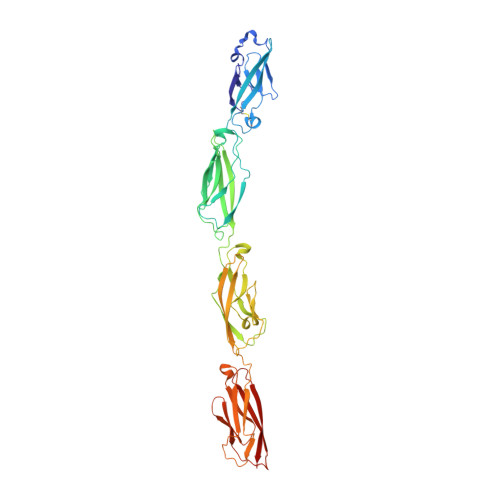
6MEQ, 6MER - PubMed Abstract:
Clustered protocadherins, a large family of paralogous proteins that play important roles in neuronal development, provide an important case study of interaction specificity in a large eukaryotic protein family. A mammalian genome has more than 50 clustered protocadherin isoforms, which have remarkable homophilic specificity for interactions between cellular surfaces. A large antiparallel dimer interface formed by the first 4 extracellular cadherin (EC) domains controls this interaction. To understand how specificity is achieved between the numerous paralogs, we used a combination of structural and computational approaches. Molecular dynamics simulations revealed that individual EC interactions are weak and undergo binding and unbinding events, but together they form a stable complex through polyvalency. Strongly evolutionarily coupled residue pairs interacted more frequently in our simulations, suggesting that sequence coevolution can inform the frequency of interaction and biochemical nature of a residue interaction. With these simulations and sequence coevolution, we generated a statistical model of interaction energy for the clustered protocadherin family that measures the contributions of all amino acid pairs at the interface. Our interaction energy model assesses specificity for all possible pairs of isoforms, recapitulating known pairings and predicting the effects of experimental changes in isoform specificity that are consistent with literature results. Our results show that sequence coevolution can be used to understand specificity determinants in a protein family and prioritize interface amino acid substitutions to reprogram specific protein-protein interactions.
- Department of Molecular and Cellular Biology, Harvard University, Cambridge, MA 02138.
Organizational Affiliation: