6MBQ

Crystal structure of Mg-free wild-type KRAS (2-166) bound to GMPPNP in the state 1 conformation


Experimental Data Snapshot

  • Method: X-RAY DIFFRACTION
  • Resolution: 1.35 Å
  • R-Value Free: 0.179 
  • R-Value Work: 0.134 
  • R-Value Observed: 0.137 

wwPDB Validation   3D Report Full Report


Ligand Structure Quality Assessment 


This is version 1.1 of the entry. See complete history


Literature

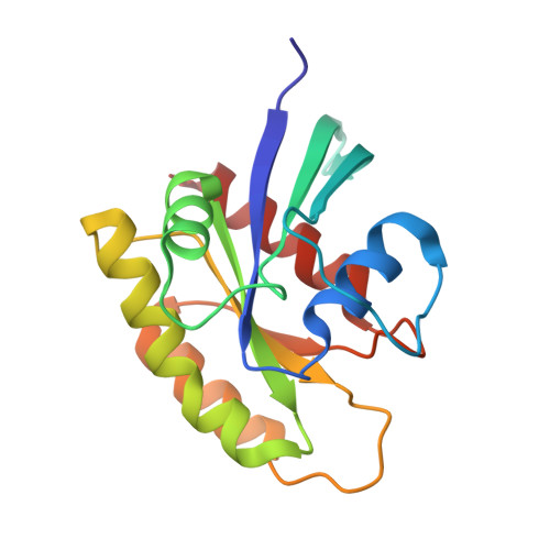
Structures of N-terminally processed KRAS provide insight into the role of N-acetylation.

Dharmaiah, S.Tran, T.H.Messing, S.Agamasu, C.Gillette, W.K.Yan, W.Waybright, T.Alexander, P.Esposito, D.Nissley, D.V.McCormick, F.Stephen, A.G.Simanshu, D.K.

(2019) Sci Rep 9: 10512-10512

  • DOI: 10.1038/s41598-019-46846-w
  • Primary Citation of Related Structures:  
    6M9W, 6MBQ, 6MBT, 6MBU, 6P0Z

  • PubMed Abstract: 
  • Although post-translational modification of the C-terminus of RAS has been studied extensively, little is known about N-terminal processing. Mass spectrometric characterization of KRAS expressed in mammalian cells showed cleavage of the initiator methionine (iMet) and N-acetylation of the nascent N-terminus ...

    Although post-translational modification of the C-terminus of RAS has been studied extensively, little is known about N-terminal processing. Mass spectrometric characterization of KRAS expressed in mammalian cells showed cleavage of the initiator methionine (iMet) and N-acetylation of the nascent N-terminus. Interestingly, structural studies on GDP- and GMPPNP-bound KRAS lacking the iMet and N-acetylation resulted in Mg 2+ -free structures of KRAS with flexible N-termini. In the Mg 2+ -free KRAS-GDP structure, the flexible N-terminus causes conformational changes in the interswitch region resulting in a fully open conformation of switch I. In the Mg 2+ -free KRAS-GMPPNP structure, the flexible N-terminus causes conformational changes around residue A59 resulting in the loss of Mg 2+ and switch I in the inactive state 1 conformation. Structural studies on N-acetylated KRAS-GDP lacking the iMet revealed the presence of Mg 2+ and a conformation of switch regions also observed in the structure of GDP-bound unprocessed KRAS with the iMet. In the absence of the iMet, the N-acetyl group interacts with the central beta-sheet and stabilizes the N-terminus and the switch regions. These results suggest there is crosstalk between the N-terminus and the Mg 2+ binding site, and that N-acetylation plays an important role by stabilizing the N-terminus of RAS upon excision of the iMet.


    Organizational Affiliation

    NCI RAS Initiative, Cancer Research Technology Program, Frederick National Laboratory for Cancer Research, Leidos Biomedical Research, Inc., Frederick, MD, 21701, USA. dhirendra.simanshu@fnlcr.nih.gov.


Macromolecules
Find similar proteins by:  (by identity cutoff)  |  3D Structure
Entity ID: 1
MoleculeChainsSequence LengthOrganismDetailsImage
GTPase KRasA167Homo sapiensMutation(s): 0 
Gene Names: KRASKRAS2RASK2
EC: 3.6.5.2
UniProt & NIH Common Fund Data Resources
Find proteins for P01116 (Homo sapiens)
Explore P01116 
Go to UniProtKB:  P01116
PHAROS:  P01116
Entity Groups  
Sequence Clusters30% Identity50% Identity70% Identity90% Identity95% Identity100% Identity
UniProt GroupP01116
Protein Feature View
Expand
  • Reference Sequence
Small Molecules
Ligands 3 Unique
IDChainsName / Formula / InChI Key2D Diagram3D Interactions
GNP (Subject of Investigation/LOI)
Query on GNP

Download Ideal Coordinates CCD File 
D [auth A]PHOSPHOAMINOPHOSPHONIC ACID-GUANYLATE ESTER
C10 H17 N6 O13 P3
UQABYHGXWYXDTK-UUOKFMHZSA-N
 Ligand Interaction
EDO
Query on EDO

Download Ideal Coordinates CCD File 
C [auth A]1,2-ETHANEDIOL
C2 H6 O2
LYCAIKOWRPUZTN-UHFFFAOYSA-N
 Ligand Interaction
NA
Query on NA

Download Ideal Coordinates CCD File 
B [auth A]SODIUM ION
Na
FKNQFGJONOIPTF-UHFFFAOYSA-N
 Ligand Interaction
Experimental Data & Validation

Experimental Data

  • Method: X-RAY DIFFRACTION
  • Resolution: 1.35 Å
  • R-Value Free: 0.179 
  • R-Value Work: 0.134 
  • R-Value Observed: 0.137 
  • Space Group: P 21 21 21
Unit Cell:
Length ( Å )Angle ( ˚ )
a = 36.02α = 90
b = 37.91β = 90
c = 99.99γ = 90
Software Package:
Software NamePurpose
XDSdata reduction
XSCALEdata scaling
PHASERphasing
PHENIXrefinement
PDB_EXTRACTdata extraction

Structure Validation

View Full Validation Report


Ligand Structure Quality Assessment 


Entry History & Funding Information

Deposition Data


Funding OrganizationLocationGrant Number
National Institutes of Health/National Cancer Institute (NIH/NCI)United StatesHHSN261200800001E

Revision History  (Full details and data files)

  • Version 1.0: 2019-07-31
    Type: Initial release
  • Version 1.1: 2019-12-04
    Changes: Author supporting evidence