6LZ7

Tetrameric structure of ZmCRY1a PHR domain


Experimental Data Snapshot

  • Method: X-RAY DIFFRACTION
  • Resolution: 3.60 Å
  • R-Value Free: 0.273 
  • R-Value Work: 0.232 
  • R-Value Observed: 0.236 

wwPDB Validation   3D Report Full Report


Ligand Structure Quality Assessment 


This is version 1.2 of the entry. See complete history


Literature

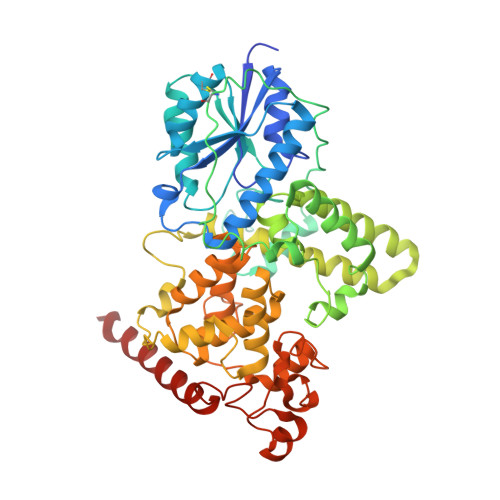
The oligomeric structures of plant cryptochromes.

Shao, K.Zhang, X.Li, X.Hao, Y.Huang, X.Ma, M.Zhang, M.Yu, F.Liu, H.Zhang, P.

(2020) Nat Struct Mol Biol 27: 480-488

  • DOI: https://doi.org/10.1038/s41594-020-0420-x
  • Primary Citation of Related Structures:  
    6LZ3, 6LZ7

  • PubMed Abstract: 

    Cryptochromes (CRYs) are a group of evolutionarily conserved flavoproteins found in many organisms. In plants, the well-studied CRY photoreceptor, activated by blue light, plays essential roles in plant growth and development. However, the mechanism of activation remains largely unknown. Here, we determined the oligomeric structures of the blue-light-perceiving PHR domain of Zea mays CRY1 and an Arabidopsis CRY2 constitutively active mutant. The structures form dimers and tetramers whose functional importance is examined in vitro and in vivo with Arabidopsis CRY2. Structure-based analysis suggests that blue light may be perceived by CRY to cause conformational changes, whose precise nature remains to be determined, leading to oligomerization that is essential for downstream signaling. This photoactivation mechanism may be widely used by plant CRYs. Our study reveals a molecular mechanism of plant CRY activation and also paves the way for design of CRY as a more efficient optical switch.


  • Organizational Affiliation

    National Key Laboratory of Plant Molecular Genetics, CAS Center for Excellence in Molecular Plant Sciences, Institute of Plant Physiology and Ecology, Shanghai Institutes for Biological Sciences, Chinese Academy of Sciences, Shanghai, China.


Macromolecules
Find similar proteins by:  (by identity cutoff)  |  3D Structure
Entity ID: 1
MoleculeChains Sequence LengthOrganismDetailsImage
Cryptochrome-1505Zea maysMutation(s): 0 
Gene Names: 100384475ZEAMMB73_Zm00001d016915
UniProt
Find proteins for A0A1D6HB67 (Zea mays)
Explore A0A1D6HB67 
Go to UniProtKB:  A0A1D6HB67
Entity Groups  
Sequence Clusters30% Identity50% Identity70% Identity90% Identity95% Identity100% Identity
UniProt GroupA0A1D6HB67
Sequence Annotations
Expand
  • Reference Sequence
Small Molecules
Ligands 1 Unique
IDChains Name / Formula / InChI Key2D Diagram3D Interactions
FAD (Subject of Investigation/LOI)
Query on FAD

Download Ideal Coordinates CCD File 
B [auth A]FLAVIN-ADENINE DINUCLEOTIDE
C27 H33 N9 O15 P2
VWWQXMAJTJZDQX-UYBVJOGSSA-N
Experimental Data & Validation

Experimental Data

  • Method: X-RAY DIFFRACTION
  • Resolution: 3.60 Å
  • R-Value Free: 0.273 
  • R-Value Work: 0.232 
  • R-Value Observed: 0.236 
  • Space Group: P 62 2 2
Unit Cell:
Length ( Å )Angle ( ˚ )
a = 154.023α = 90
b = 154.023β = 90
c = 141.072γ = 120
Software Package:
Software NamePurpose
PHENIXrefinement
PHENIXrefinement
HKL-3000data collection
HKL-3000data scaling
PHENIXphasing
PHENIXmodel building

Structure Validation

View Full Validation Report


Ligand Structure Quality Assessment 


Entry History 

Deposition Data

Revision History  (Full details and data files)

  • Version 1.0: 2020-05-13
    Type: Initial release
  • Version 1.1: 2020-05-27
    Changes: Database references
  • Version 1.2: 2023-11-29
    Changes: Data collection, Database references, Refinement description