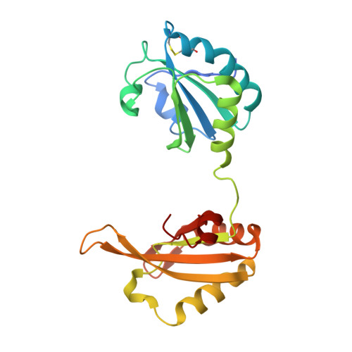
6LUR

Human PUF60 UHM domain (thioredoxin fusion) in complex with a small molecule binder


Experimental Data Snapshot

  • Method: X-RAY DIFFRACTION
  • Resolution: 2.00 Å
  • R-Value Free: 0.253 
  • R-Value Work: 0.201 
  • R-Value Observed: 0.203 

wwPDB Validation   3D Report Full Report


Ligand Structure Quality Assessment 


This is version 1.2 of the entry. See complete history


Literature

Revisiting biomolecular NMR spectroscopy for promoting small-molecule drug discovery.

Hanzawa, H.Shimada, T.Takahashi, M.Takahashi, H.

(2020) J Biomol NMR 74: 501-508

  • DOI: https://doi.org/10.1007/s10858-020-00314-0
  • Primary Citation of Related Structures:  
    6LUR

  • PubMed Abstract: 
  • Recently, there has been increasing interest in new modalities such as therapeutic antibodies and gene therapy at a number of pharmaceutical companies. Moreover, in small-molecule drug discovery at such companies, efforts have focused on hard-to-drug targets such as inhibiting protein-protein interactions ...

    Recently, there has been increasing interest in new modalities such as therapeutic antibodies and gene therapy at a number of pharmaceutical companies. Moreover, in small-molecule drug discovery at such companies, efforts have focused on hard-to-drug targets such as inhibiting protein-protein interactions. Biomolecular NMR spectroscopy has been used in drug discovery in a variety of ways, such as for the reliable detection of binding and providing three-dimensional structural information for structure-based drug design. The advantages of using NMR spectroscopy have been known for decades (Jahnke in J Biomol NMR 39:87-90, (2007); Gossert and Jahnke in Prog Nucl Magn Reson Spectrosc 97:82-125, (2016)). For tackling hard-to-drug targets and increasing the success in discovering drug molecules, in-depth analysis of drug-target protein interactions performed by biophysical methods will be more and more essential. Here, we review the advantages of NMR spectroscopy as a key technology of biophysical methods and also discuss issues such as using cutting-edge NMR spectrometers and increasing the demand of utilizing conformational dynamics information for promoting small-molecule drug discovery.


    Organizational Affiliation

    Graduate School of Medical Life Science, Yokohama City University, 1-7-29 Suehiro-cho, Tsurumi-ku, Yokohama, 230-0045, Japan.


Macromolecules
Find similar proteins by:  (by identity cutoff)  |  3D Structure
Entity ID: 1
MoleculeChainsSequence LengthOrganismDetailsImage
Thioredoxin 1,Poly(U)-binding-splicing factor PUF60
A, B, C, D, E
A, B, C, D, E, F, G, H
222Escherichia coli K-12Homo sapiens
This entity is chimeric
Mutation(s): 0 
Gene Names: trxAfipAtsnCb3781JW5856PUF60FIRROBPISIAHBP1
UniProt & NIH Common Fund Data Resources
Find proteins for P0AA25 (Escherichia coli (strain K12))
Explore P0AA25 
Go to UniProtKB:  P0AA25
Find proteins for Q9UHX1 (Homo sapiens)
Explore Q9UHX1 
Go to UniProtKB:  Q9UHX1
PHAROS:  Q9UHX1
Entity Groups  
Sequence Clusters30% Identity50% Identity70% Identity90% Identity95% Identity100% Identity
UniProt GroupsP0AA25Q9UHX1
Protein Feature View
Expand
  • Reference Sequence
Small Molecules
Binding Affinity Annotations 
IDSourceBinding Affinity
EVU Binding MOAD:  6LUR Kd: 1.95e+5 (nM) from 1 assay(s)
Experimental Data & Validation

Experimental Data

  • Method: X-RAY DIFFRACTION
  • Resolution: 2.00 Å
  • R-Value Free: 0.253 
  • R-Value Work: 0.201 
  • R-Value Observed: 0.203 
  • Space Group: P 21 21 21
Unit Cell:
Length ( Å )Angle ( ˚ )
a = 77.019α = 90
b = 90.59β = 90
c = 288.928γ = 90
Software Package:
Software NamePurpose
XSCALEdata scaling
REFMACrefinement
PDB_EXTRACTdata extraction
XDSdata reduction
PHASERphasing

Structure Validation

View Full Validation Report


Ligand Structure Quality Assessment 


Entry History 

Deposition Data

Revision History  (Full details and data files)

  • Version 1.0: 2020-04-29
    Type: Initial release
  • Version 1.1: 2020-05-06
    Changes: Database references
  • Version 1.2: 2020-12-09
    Changes: Database references