Haloperidol bound D2dopamine receptor structure inspired the discovery of subtype selective ligands.
Fan, L., Tan, L., Chen, Z., Qi, J., Nie, F., Luo, Z., Cheng, J., Wang, S.(2020) Nat Commun 11: 1074-1074
- PubMed: 32103023 Search on PubMedSearch on PubMed Central
- DOI: https://doi.org/10.1038/s41467-020-14884-y
- Primary Citation Related Structures:
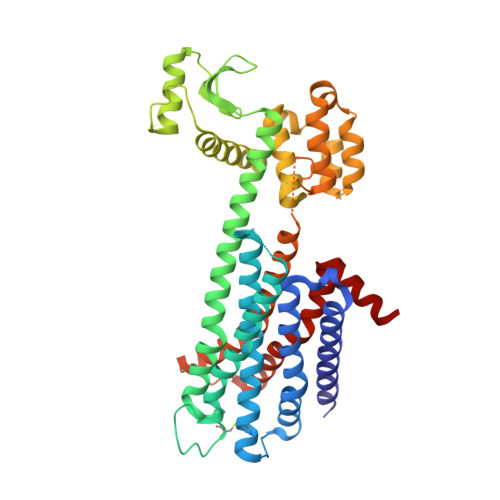
6LUQ - PubMed Abstract:
The D 2 dopamine receptor (DRD2) is one of the most well-established therapeutic targets for neuropsychiatric and endocrine disorders. Most clinically approved and investigational drugs that target this receptor are known to be subfamily-selective for all three D 2 -like receptors, rather than subtype-selective for only DRD2. Here, we report the crystal structure of DRD2 bound to the most commonly used antipsychotic drug, haloperidol. The structures suggest an extended binding pocket for DRD2 that distinguishes it from other D 2 -like subtypes. A detailed analysis of the structures illuminates key structural determinants essential for DRD2 activation and subtype selectivity. A structure-based and mechanism-driven screening combined with a lead optimization approach yield DRD2 highly selective agonists, which could be used as chemical probes for studying the physiological and pathological functions of DRD2 as well as promising therapeutic leads devoid of promiscuity.
- State Key Laboratory of Molecular Biology, CAS Center for Excellence in Molecular Cell Science, Shanghai Institute of Biochemistry and Cell Biology, Chinese Academy of Sciences, University of Chinese Academy of Sciences, 320 Yueyang Road, 200031, Shanghai, China.
Organizational Affiliation: