6LPG

human VASH1-SVBP complex


Experimental Data Snapshot

  • Method: X-RAY DIFFRACTION
  • Resolution: 2.30 Å
  • R-Value Free: 0.236 
  • R-Value Work: 0.190 
  • R-Value Observed: 0.194 

Starting Model: experimental
View more details

wwPDB Validation   3D Report Full Report


Ligand Structure Quality Assessment 


This is version 1.1 of the entry. See complete history


Literature

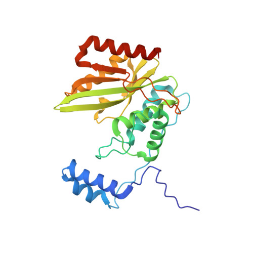
The crystal structure of the tetrameric human vasohibin-1-SVBP complex reveals a variable arm region within the structural core.

Ikeda, A.Urata, S.Ando, T.Suzuki, Y.Sato, Y.Nishino, T.

(2020) Acta Crystallogr D Struct Biol 76: 993-1000

  • DOI: https://doi.org/10.1107/S2059798320011298
  • Primary Citation of Related Structures:  
    6LPG

  • PubMed Abstract: 

    Vasohibins regulate angiogenesis, tumor growth, metastasis and neuronal differentiation. They form a complex with small vasohibin-binding protein (SVBP) and show tubulin tyrosine carboxypeptidase activity. Recent crystal structure determinations of vasohibin-SVBP complexes have provided a molecular basis for complex formation, substrate binding and catalytic activity. However, the regulatory mechanism and dynamics of the complex remain elusive. Here, the crystal structure of the VASH1-SVBP complex and a molecular-dynamics simulation study are reported. The overall structure of the complex was similar to previously reported structures. Importantly, however, the structure revealed a domain-swapped heterotetramer that was formed between twofold symmetry-related molecules. This heterotetramerization was stabilized by the mutual exchange of ten conserved N-terminal residues from the VASH1 structural core, which was intramolecular in other structures. Interestingly, a comparison of this region with previously reported structures revealed that the patterns of hydrogen bonding and hydrophobic interactions vary. In the molecular-dynamics simulations, differences were found between the heterotetramer and heterodimer, where the fluctuation of the N-terminal region in the heterotetramer was suppressed. Thus, heterotetramer formation and flexibility of the N-terminal region may be important for enzyme activity and regulation.


  • Organizational Affiliation

    Department of Biological Science and Technology, Tokyo University of Science, 6-3-1 Niijyuku, Katsushika-ku, Tokyo 125-8585, Japan.


Macromolecules
Find similar proteins by:  (by identity cutoff)  |  3D Structure
Entity ID: 1
MoleculeChains Sequence LengthOrganismDetailsImage
Tubulinyl-Tyr carboxypeptidase 1256Homo sapiensMutation(s): 0 
Gene Names: VASH1KIAA1036VASH
EC: 3.4.17.17
UniProt & NIH Common Fund Data Resources
Find proteins for Q7L8A9 (Homo sapiens)
Explore Q7L8A9 
Go to UniProtKB:  Q7L8A9
PHAROS:  Q7L8A9
GTEx:  ENSG00000071246 
Entity Groups  
Sequence Clusters30% Identity50% Identity70% Identity90% Identity95% Identity100% Identity
UniProt GroupQ7L8A9
Sequence Annotations
Expand
  • Reference Sequence
Find similar proteins by:  (by identity cutoff)  |  3D Structure
Entity ID: 2
MoleculeChains Sequence LengthOrganismDetailsImage
Small vasohibin-binding protein67Homo sapiensMutation(s): 0 
Gene Names: SVBPCCDC23
UniProt & NIH Common Fund Data Resources
Find proteins for Q8N300 (Homo sapiens)
Explore Q8N300 
Go to UniProtKB:  Q8N300
PHAROS:  Q8N300
GTEx:  ENSG00000177868 
Entity Groups  
Sequence Clusters30% Identity50% Identity70% Identity90% Identity95% Identity100% Identity
UniProt GroupQ8N300
Sequence Annotations
Expand
  • Reference Sequence
Experimental Data & Validation

Experimental Data

  • Method: X-RAY DIFFRACTION
  • Resolution: 2.30 Å
  • R-Value Free: 0.236 
  • R-Value Work: 0.190 
  • R-Value Observed: 0.194 
  • Space Group: P 61 2 2
Unit Cell:
Length ( Å )Angle ( ˚ )
a = 71.854α = 90
b = 71.854β = 90
c = 215.811γ = 120
Software Package:
Software NamePurpose
PHENIXrefinement
XDSdata reduction
pointlessdata scaling
PHASERphasing

Structure Validation

View Full Validation Report


Ligand Structure Quality Assessment 


Entry History & Funding Information

Deposition Data


Funding OrganizationLocationGrant Number
Japan Society for the Promotion of ScienceJapan16K07279

Revision History  (Full details and data files)

  • Version 1.0: 2020-10-21
    Type: Initial release
  • Version 1.1: 2023-11-29
    Changes: Advisory, Data collection, Database references, Refinement description