6LKP | pdb_00006lkp

Crystal structure of Dps1 from the thermophilic non-heterocystous filamentous cyanobacterium Thermoleptolyngbya sp. O-77


Experimental Data Snapshot

  • Method: X-RAY DIFFRACTION
  • Resolution: 2.90 Å
  • R-Value Free: 
    0.315 (Depositor), 0.313 (DCC) 
  • R-Value Work: 
    0.253 (Depositor), 0.254 (DCC) 
  • R-Value Observed: 
    0.257 (Depositor) 

Starting Model: experimental
View more details

wwPDB Validation 3D Report Full Report

Validation slider image for 6LKP

This is version 2.2 of the entry. See complete history

Literature

Biochemical and structural characterization of a thermostable Dps protein with His-type ferroxidase centers and outer metal-binding sites.

Minato, T.Teramoto, T.Kakuta, Y.Ogo, S.Yoon, K.S.

(2020) FEBS Open Bio 10: 1219-1229

  • DOI: https://doi.org/10.1002/2211-5463.12837
  • Primary Citation Related Structures: 
    6LKP

  • PubMed Abstract: 

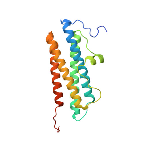
    The DNA-binding protein from starved cells (Dps) is found in a wide range of microorganisms, and it has been well characterized. However, little is known about Dps proteins from nonheterocystous filamentous cyanobacteria. In this study, a Dps protein from the thermophilic nonheterocystous filamentous cyanobacterium Thermoleptolyngbya sp. O-77 (TlDps1) was purified and characterized. PAGE and CD analyses of TlDps1 demonstrated that it had higher thermostability than previously reported Dps proteins. X-ray crystallographic analysis revealed that TlDps1 possessed His-type ferroxidase centers within the cavity and unique metal-binding sites located on the surface of the protein, which presumably contributed to its exceedingly high thermostability.


  • Organizational Affiliation
    • Department of Chemistry and Biochemistry, Graduate School of Engineering, Kyushu University, Fukuoka, Japan.

Macromolecule Content 

  • Total Structure Weight: 125.48 kDa 
  • Atom Count: 7,992 
  • Modeled Residue Count: 1,001 
  • Deposited Residue Count: 1,104 
  • Unique protein chains: 1

Macromolecules

Find similar proteins by:|  3D Structure
Entity ID: 1
MoleculeChains  Sequence LengthOrganismDetailsImage
DNA protection during starvation protein
A, B, C, D, E
A, B, C, D, E, F
184Leptolyngbya sp. O-77Mutation(s): 0 
EC: 1.16

Small Molecules

Ligands 2 Unique
IDChains Name / Formula / InChI Key2D Diagram3D Interactions
ZN
(Subject of Investigation/LOI)

Query on ZN


Download:Ideal Coordinates CCD File
BA [auth F]
G [auth A]
H [auth A]
K [auth A]
L [auth B]
BA [auth F],
G [auth A],
H [auth A],
K [auth A],
L [auth B],
O [auth B],
P [auth B],
S [auth C],
T [auth D],
U [auth D],
X [auth D],
Y [auth E]
ZINC ION
Zn
PTFCDOFLOPIGGS-UHFFFAOYSA-N
FE
(Subject of Investigation/LOI)

Query on FE


Download:Ideal Coordinates CCD File
AA [auth E]
CA [auth F]
DA [auth F]
I [auth A]
J [auth A]
AA [auth E],
CA [auth F],
DA [auth F],
I [auth A],
J [auth A],
M [auth B],
N [auth B],
Q [auth C],
R [auth C],
V [auth D],
W [auth D],
Z [auth E]
FE (III) ION
Fe
VTLYFUHAOXGGBS-UHFFFAOYSA-N

Experimental Data & Validation

Experimental Data

  • Method: X-RAY DIFFRACTION
  • Resolution: 2.90 Å
  • R-Value Free:  0.315 (Depositor), 0.313 (DCC) 
  • R-Value Work:  0.253 (Depositor), 0.254 (DCC) 
  • R-Value Observed: 0.257 (Depositor) 
Space Group: P 43 2 2
Unit Cell:
Length ( Å )Angle ( ˚ )
a = 92.787α = 90
b = 92.787β = 90
c = 261.12γ = 90
Software Package:
Software NamePurpose
Cootmodel building
PHENIXrefinement
XDSdata reduction
XDSdata scaling
PHASERphasing

Structure Validation

View Full Validation Report


Entry History 

& Funding Information

Deposition Data


Funding OrganizationLocationGrant Number
Japan Society for the Promotion of Science (JSPS)JapanJPMJCR18R2

Revision History  (Full details and data files)

  • Version 1.0: 2020-03-25
    Type: Initial release
  • Version 2.0: 2020-04-01
    Changes: Atomic model, Data collection, Derived calculations
  • Version 2.1: 2020-09-30
    Changes: Database references, Derived calculations
  • Version 2.2: 2023-11-22
    Changes: Data collection, Database references, Refinement description