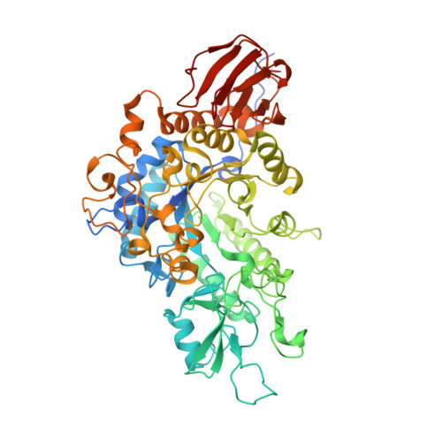
6LGI

Bombyx mori GH13 sucrose hydrolase mutant E322Q covalent intermediate complexed with fructose


Experimental Data Snapshot

  • Method: X-RAY DIFFRACTION
  • Resolution: 1.60 Å
  • R-Value Free: 0.207 
  • R-Value Work: 0.178 
  • R-Value Observed: 0.179 

Starting Model: experimental
View more details

wwPDB Validation   3D Report Full Report


Ligand Structure Quality Assessment 


This is version 1.3 of the entry. See complete history


Literature

Structure-function analysis of silkworm sucrose hydrolase uncovers the mechanism of substrate specificity in GH13 subfamily 17exo-alpha-glucosidases.

Miyazaki, T.Park, E.Y.

(2020) J Biol Chem 295: 8784-8797

  • DOI: https://doi.org/10.1074/jbc.RA120.013595
  • Primary Citation of Related Structures:  
    6LGA, 6LGB, 6LGC, 6LGD, 6LGE, 6LGF, 6LGG, 6LGH, 6LGI

  • PubMed Abstract: 

    The domestic silkworm Bombyx mori expresses two sucrose-hydrolyzing enzymes, BmSUH and BmSUC1, belonging to glycoside hydrolase family 13 subfamily 17 (GH13_17) and GH32, respectively. BmSUH has little activity on maltooligosaccharides, whereas other insect GH13_17 α-glucosidases are active on sucrose and maltooligosaccharides. Little is currently known about the structural mechanisms and substrate specificity of GH13_17 enzymes. In this study, we examined the crystal structures of BmSUH without ligands; in complexes with substrates, products, and inhibitors; and complexed with its covalent intermediate at 1.60-1.85 Å resolutions. These structures revealed that the conformations of amino acid residues around subsite -1 are notably different at each step of the hydrolytic reaction. Such changes have not been previously reported among GH13 enzymes, including exo - and endo -acting hydrolases, such as α-glucosidases and α-amylases. Amino acid residues at subsite +1 are not conserved in BmSUH and other GH13_17 α-glucosidases, but subsite -1 residues are absolutely conserved. Substitutions in three subsite +1 residues, Gln 191 , Tyr 251 , and Glu 440 , decreased sucrose hydrolysis and increased maltase activity of BmSUH, indicating that these residues are key for determining its substrate specificity. These results provide detailed insights into structure-function relationships in GH13 enzymes and into the molecular evolution of insect GH13_17 α-glucosidases.


  • Organizational Affiliation

    Green Chemistry Research Division, Research Institute of Green Science and Technology, Shizuoka University, Shizuoka, Japan. Electronic address: miyazaki.takatsugu@shizuoka.ac.jp.


Macromolecules
Find similar proteins by:  (by identity cutoff)  |  3D Structure
Entity ID: 1
MoleculeChains Sequence LengthOrganismDetailsImage
Sucrose hydrolase
A, B
598Bombyx moriMutation(s): 1 
Gene Names: BmSuh
UniProt
Find proteins for A0A077JI83 (Bombyx mori)
Explore A0A077JI83 
Go to UniProtKB:  A0A077JI83
Entity Groups  
Sequence Clusters30% Identity50% Identity70% Identity90% Identity95% Identity100% Identity
UniProt GroupA0A077JI83
Sequence Annotations
Expand
  • Reference Sequence
Small Molecules
Ligands 6 Unique
IDChains Name / Formula / InChI Key2D Diagram3D Interactions
BDF (Subject of Investigation/LOI)
Query on BDF

Download Ideal Coordinates CCD File 
H [auth A],
P [auth B]
beta-D-fructopyranose
C6 H12 O6
LKDRXBCSQODPBY-ARQDHWQXSA-N
FRU (Subject of Investigation/LOI)
Query on FRU

Download Ideal Coordinates CCD File 
G [auth A],
O [auth B]
beta-D-fructofuranose
C6 H12 O6
RFSUNEUAIZKAJO-ARQDHWQXSA-N
BGC (Subject of Investigation/LOI)
Query on BGC

Download Ideal Coordinates CCD File 
F [auth A],
N [auth B]
beta-D-glucopyranose
C6 H12 O6
WQZGKKKJIJFFOK-VFUOTHLCSA-N
ACT
Query on ACT

Download Ideal Coordinates CCD File 
I [auth A],
Q [auth B]
ACETATE ION
C2 H3 O2
QTBSBXVTEAMEQO-UHFFFAOYSA-M
CA (Subject of Investigation/LOI)
Query on CA

Download Ideal Coordinates CCD File 
D [auth A],
L [auth B]
CALCIUM ION
Ca
BHPQYMZQTOCNFJ-UHFFFAOYSA-N
MG (Subject of Investigation/LOI)
Query on MG

Download Ideal Coordinates CCD File 
C [auth A],
E [auth A],
J [auth A],
K [auth B],
M [auth B]
MAGNESIUM ION
Mg
JLVVSXFLKOJNIY-UHFFFAOYSA-N
Experimental Data & Validation

Experimental Data

  • Method: X-RAY DIFFRACTION
  • Resolution: 1.60 Å
  • R-Value Free: 0.207 
  • R-Value Work: 0.178 
  • R-Value Observed: 0.179 
  • Space Group: P 21 21 21
Unit Cell:
Length ( Å )Angle ( ˚ )
a = 65.405α = 90
b = 147.055β = 90
c = 153.544γ = 90
Software Package:
Software NamePurpose
REFMACrefinement
PDB_EXTRACTdata extraction
XDSdata reduction
XDSdata scaling
MOLREPphasing

Structure Validation

View Full Validation Report


Ligand Structure Quality Assessment 


Entry History & Funding Information

Deposition Data

  • Released Date: 2020-05-20 
  • Deposition Author(s): Miyazaki, T.

Funding OrganizationLocationGrant Number
Japan Society for the Promotion of ScienceJapan19K15748

Revision History  (Full details and data files)

  • Version 1.0: 2020-05-20
    Type: Initial release
  • Version 1.1: 2020-07-08
    Changes: Database references
  • Version 1.2: 2020-07-29
    Type: Remediation
    Reason: Carbohydrate remediation
    Changes: Data collection, Derived calculations, Structure summary
  • Version 1.3: 2023-11-22
    Changes: Data collection, Database references, Refinement description, Structure summary