6LFJ

Crystal structure of mouse DCAR2 CRD domain complex with IPM2


Experimental Data Snapshot

  • Method: X-RAY DIFFRACTION
  • Resolution: 1.84 Å
  • R-Value Free: 0.198 
  • R-Value Work: 0.177 
  • R-Value Observed: 0.178 

Starting Model: experimental
View more details

wwPDB Validation   3D Report Full Report


Ligand Structure Quality Assessment 


This is version 1.3 of the entry. See complete history


Literature

Structural insight into the recognition of pathogen-derived phosphoglycolipids by C-type lectin receptor DCAR.

Omahdi, Z.Horikawa, Y.Nagae, M.Toyonaga, K.Imamura, A.Takato, K.Teramoto, T.Ishida, H.Kakuta, Y.Yamasaki, S.

(2020) J Biol Chem 295: 5807-5817

  • DOI: https://doi.org/10.1074/jbc.RA120.012491
  • Primary Citation of Related Structures:  
    6KZR, 6LFJ, 6LKR

  • PubMed Abstract: 

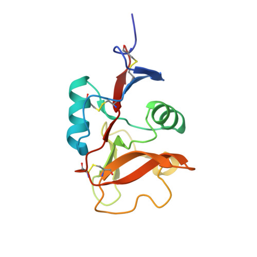
    The C-type lectin receptors (CLRs) form a family of pattern recognition receptors that recognize numerous pathogens, such as bacteria and fungi, and trigger innate immune responses. The extracellular carbohydrate-recognition domain (CRD) of CLRs forms a globular structure that can coordinate a Ca 2+ ion, allowing receptor interactions with sugar-containing ligands. Although well-conserved, the CRD fold can also display differences that directly affect the specificity of the receptors for their ligands. Here, we report crystal structures at 1.8-2.3 Å resolutions of the CRD of murine dendritic cell-immunoactivating receptor (DCAR, or Clec4b1 ), the CLR that binds phosphoglycolipids such as acylated phosphatidyl- myo -inositol mannosides (AcPIMs) of mycobacteria. Using mutagenesis analysis, we identified critical residues, Ala 136 and Gln 198 , on the surface surrounding the ligand-binding site of DCAR, as well as an atypical Ca 2+ -binding motif (Glu-Pro-Ser/EPS 168-170 ). By chemically synthesizing a water-soluble ligand analog, inositol-monophosphate dimannose (IPM2), we confirmed the direct interaction of DCAR with the polar moiety of AcPIMs by biolayer interferometry and co-crystallization approaches. We also observed a hydrophobic groove extending from the ligand-binding site that is in a suitable position to interact with the lipid portion of whole AcPIMs. These results suggest that the hydroxyl group-binding ability and hydrophobic groove of DCAR mediate its specific binding to pathogen-derived phosphoglycolipids such as mycobacterial AcPIMs.


  • Organizational Affiliation

    Department of Molecular Immunology, Research Institute for Microbial Diseases, Osaka University, Suita 565-0871, Japan; Laboratory of Molecular Immunology, Immunology Frontier Research Center (IFReC), Osaka University, Suita 565-0871, Japan; Division of Molecular Immunology, Medical Institute of Bioregulation, Kyushu University, Fukuoka 812-8582, Japan.


Macromolecules
Find similar proteins by:  (by identity cutoff)  |  3D Structure
Entity ID: 1
MoleculeChains Sequence LengthOrganismDetailsImage
C-type lectin domain family 4, member b1
A, B
136Mus musculusMutation(s): 0 
Gene Names: Clec4b1Clec4bDcar
UniProt
Find proteins for Q9D8Q7 (Mus musculus)
Explore Q9D8Q7 
Go to UniProtKB:  Q9D8Q7
Entity Groups  
Sequence Clusters30% Identity50% Identity70% Identity90% Identity95% Identity100% Identity
UniProt GroupQ9D8Q7
Sequence Annotations
Expand
  • Reference Sequence
Small Molecules
Ligands 4 Unique
IDChains Name / Formula / InChI Key2D Diagram3D Interactions
B3P
Query on B3P

Download Ideal Coordinates CCD File 
D [auth A]2-[3-(2-HYDROXY-1,1-DIHYDROXYMETHYL-ETHYLAMINO)-PROPYLAMINO]-2-HYDROXYMETHYL-PROPANE-1,3-DIOL
C11 H26 N2 O6
HHKZCCWKTZRCCL-UHFFFAOYSA-N
IPD
Query on IPD

Download Ideal Coordinates CCD File 
F [auth A],
I [auth B]
D-MYO-INOSITOL-1-PHOSPHATE
C6 H11 O9 P
INAPMGSXUVUWAF-UOTPTPDRSA-L
MAN
Query on MAN

Download Ideal Coordinates CCD File 
E [auth A],
G [auth A],
J [auth B]
alpha-D-mannopyranose
C6 H12 O6
WQZGKKKJIJFFOK-PQMKYFCFSA-N
CA
Query on CA

Download Ideal Coordinates CCD File 
C [auth A],
H [auth B]
CALCIUM ION
Ca
BHPQYMZQTOCNFJ-UHFFFAOYSA-N
Experimental Data & Validation

Experimental Data

  • Method: X-RAY DIFFRACTION
  • Resolution: 1.84 Å
  • R-Value Free: 0.198 
  • R-Value Work: 0.177 
  • R-Value Observed: 0.178 
  • Space Group: P 21 21 21
Unit Cell:
Length ( Å )Angle ( ˚ )
a = 65.689α = 90
b = 72.564β = 90
c = 100.469γ = 90
Software Package:
Software NamePurpose
PHENIXrefinement
XDSdata reduction
XDSdata scaling
MOLREPphasing

Structure Validation

View Full Validation Report


Ligand Structure Quality Assessment 


Entry History 

Deposition Data

Revision History  (Full details and data files)

  • Version 1.0: 2020-03-25
    Type: Initial release
  • Version 1.1: 2020-05-06
    Changes: Database references
  • Version 1.2: 2020-07-29
    Type: Remediation
    Reason: Carbohydrate remediation
    Changes: Data collection, Derived calculations, Structure summary
  • Version 1.3: 2023-11-22
    Changes: Data collection, Database references, Derived calculations, Refinement description, Structure summary