6L2W | pdb_00006l2w

Crystal structure of a novel fold protein Gp72 from the freshwater cyanophage Mic1


Experimental Data Snapshot

  • Method: X-RAY DIFFRACTION
  • Resolution: 2.29 Å
  • R-Value Free: 
    0.238 (Depositor), 0.244 (DCC) 
  • R-Value Work: 
    0.199 (Depositor), 0.207 (DCC) 
  • R-Value Observed: 
    0.201 (Depositor) 

wwPDB Validation 3D Report Full Report

Validation slider image for 6L2W

This is version 1.2 of the entry. See complete history

Literature

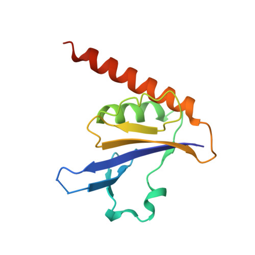
Crystal structure of a novel fold protein Gp72 from the freshwater cyanophage Mic1.

Wang, Y.Jin, H.Yang, F.Jiang, Y.L.Zhao, Y.Y.Chen, Z.P.Li, W.F.Chen, Y.Zhou, C.Z.Li, Q.

(2020) Proteins 88: 1226-1232

  • DOI: https://doi.org/10.1002/prot.25896
  • Primary Citation Related Structures: 
    6L2W

  • PubMed Abstract: 

    Cyanophages, widespread in aquatic systems, are a class of viruses that specifically infect cyanobacteria. Though they play important roles in modulating the homeostasis of cyanobacterial populations, little is known about the freshwater cyanophages, especially those hypothetical proteins of unknown function. Mic1 is a freshwater siphocyanophage isolated from the Lake Chaohu. It encodes three hypothetical proteins Gp65, Gp66, and Gp72, which share an identity of 61.6% to 83%. However, we find these three homologous proteins differ from each other in oligomeric state. Moreover, we solve the crystal structure of Gp72 at 2.3 Å, which represents a novel fold in the α + β class. Structural analyses combined with redox assays enable us to propose a model of disulfide bond mediated oligomerization for Gp72. Altogether, these findings provide structural and biochemical basis for further investigations on the freshwater cyanophage Mic1.


  • Organizational Affiliation
    • Hefei National Laboratory for Physical Sciences at the Microscale and School of Life Sciences, University of Science and Technology of China, Hefei, Anhui, China.

Macromolecule Content 

  • Total Structure Weight: 29.27 kDa 
  • Atom Count: 1,976 
  • Modeled Residue Count: 234 
  • Deposited Residue Count: 248 
  • Unique protein chains: 1

Macromolecules

Find similar proteins by:|  3D Structure
Entity ID: 1
MoleculeChains  Sequence LengthOrganismDetailsImage
freshwater cyanophage protein
A, B
124Microcystis phage Mic1Mutation(s): 0 
UniProt
Find proteins for A0ACD6BAA9 (Microcystis phage Mic1)
Explore A0ACD6BAA9 
Go to UniProtKB:  A0ACD6BAA9
Entity Groups
Sequence Clusters30% Identity50% Identity70% Identity90% Identity95% Identity100% Identity
UniProt GroupA0ACD6BAA9
Sequence Annotations
Expand
Reference Sequence

Experimental Data & Validation

Experimental Data

  • Method: X-RAY DIFFRACTION
  • Resolution: 2.29 Å
  • R-Value Free:  0.238 (Depositor), 0.244 (DCC) 
  • R-Value Work:  0.199 (Depositor), 0.207 (DCC) 
  • R-Value Observed: 0.201 (Depositor) 
Space Group: C 1 2 1
Unit Cell:
Length ( Å )Angle ( ˚ )
a = 85.702α = 90
b = 45.534β = 94.29
c = 67.368γ = 90
Software Package:
Software NamePurpose
REFMACrefinement
HKL-2000data reduction
HKL-2000data scaling
AutoSolphasing

Structure Validation

View Full Validation Report


Entry History 

& Funding Information

Deposition Data


Funding OrganizationLocationGrant Number
National Natural Science Foundation of ChinaChina31630001
National Natural Science Foundation of ChinaChina31621002
Ministry of Science and Technology (China)China2018YFA0903100
Ministry of Science and Technology (China)China2016YFA0400900
Chinese Academy of SciencesChina--

Revision History  (Full details and data files)

  • Version 1.0: 2020-05-13
    Type: Initial release
  • Version 1.1: 2020-08-12
    Changes: Database references
  • Version 1.2: 2024-03-27
    Changes: Data collection, Database references