


Ultrafast Photoinduced Electron Transfer in a Photosensitizer Protein
Zheng, D., Tao, M., Yu, L.J., Liu, X., Xia, A., Wang, J.(2021) CC Chem 3: 1580-1586
Experimental Data Snapshot
Starting Model: experimental
View more details
wwPDB Validation 3D Report Full Report

Entity ID: 1 | |||||
|---|---|---|---|---|---|
| Molecule | Chains | Sequence Length | Organism | Details | Image |
| Yellow fluorescent protein | 238 | Aequorea victoria | Mutation(s): 15 Gene Names: yfp | 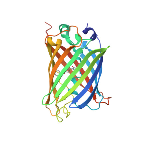 | |
UniProt | |||||
Entity Groups | |||||
| Sequence Clusters | 30% Identity50% Identity70% Identity90% Identity95% Identity100% Identity | ||||
| UniProt Group | A0A059PIR9 | ||||
Sequence AnnotationsExpand | |||||
Reference Sequence | |||||
| Modified Residues 1 Unique | |||||
|---|---|---|---|---|---|
| ID | Chains | Type | Formula | 2D Diagram | Parent |
| BF6 Query on BF6 | A | L-PEPTIDE LINKING | C20 H17 N3 O4 |  | GLY, TYR, GLY |
| Length ( Å ) | Angle ( ˚ ) |
|---|---|
| a = 51.604 | α = 90 |
| b = 51.604 | β = 90 |
| c = 179.053 | γ = 90 |
| Software Name | Purpose |
|---|---|
| HKL-2000 | data scaling |
| PHENIX | refinement |
| PDB_EXTRACT | data extraction |
| HKL-3000 | data reduction |
| PHASER | phasing |


